Introduction

Fracture Nomenclature for Distal Radius Fractures

Hand Surgery Resource’s Diagnostic Guides describe fractures by the anatomical name of the fractured bone and then characterize the fracture by the Acronym:
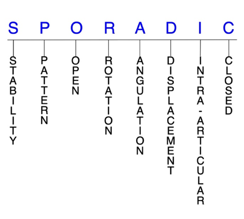
In addition, anatomically named fractures are often also identified by specific eponyms or other special features.

For the Distal Radius, the historical and specifically named fractures include:

Colles’ Fracture

Smith Fracture

Barton Fracture

Reverse Barton Fracture

Distal ulna fracture

By selecting the name (diagnosis), you will be linked to the introduction section of this Diagnostic Guide dedicated to the selected fracture eponym.


Distal radius fractures account for 12-17% of all fractures, which makes them the most common upper extremity fracture. These injuries occur most frequently in two age groups: in children and adolescents due to high-energy trauma, and in older patients with osteopenia or osteoporosis after low-to-moderate energy injuries. Despite the high prevalence of distal radius fractures and the continued emergence of new treatment options and techniques, universal consensus on the optimal management strategy is lacking. Many distal radius fractures—especially closed, stable, nondisplaced injuries—can effectively be treated conservatively with closed reduction and immobilization. Surgery is often required for open and severely displaced fractures, and for those that fail conservative treatment.1-6,26-30

Definitions

  • A distal radius fracture is a disruption of the mechanical integrity of the distal radius.
  • A distal radius fracture produces a discontinuity in the distal radius contours that can be complete or incomplete.
  • A distal radius fracture is caused by a direct force that exceeds the breaking point of the bone.

Hand Surgery Resource’s Fracture Description and Characterization Acronym

SPORADIC

S – Stability; P – Pattern; O – Open; R – Rotation; A – Angulation; D – Displacement; I – Intra-articular; C – Closed


S - Stability (stable or unstable)

  • Universally accepted definitions of clinical fracture stability is not well defined in the literature.7-9
  • Stable: fracture fragment pattern is generally nondisplaced or minimally displaced. It does not require reduction, and the fracture fragments’ alignment is maintained by with simple splinting or casting. However, most definitions define a stable fracture as one that will maintain anatomical alignment after a simple closed reduction and splinting. Some authors add that stable fractures remain aligned, even when adjacent joints are put to a partial range of motion (ROM).
  • Unstable: will not remain anatomically or nearly anatomically aligned after a successful closed reduction and immobilization. Typically unstable distal radius fractures have significant deformity with comminution, displacement, angulation, and/or shortening.
    • A distal radius fracture is further defined as unstable if it is unable to resist displacement after being anatomically reduced.10

P - Pattern

  • Distal radial styloid
  • Distal dorsal medial fragment
  • Distal volar medial fragment
  • Distal radial shaft

O - Open

  • Open: a wound connects the external environment to the fracture site. The wound provides a pathway for bacteria to reach and infect the fracture site. As a result, there is always a risk for chronic osteomyelitis. Therefore, open fractures of the distal radius require antibiotics with surgical irrigation and wound debridement.7,11,12

R - Rotation

  • Distal radius fracture deformity can be caused by proximal rotation of the fracture fragment in relation to the distal fracture fragment.
  • Degree of malrotation of the fracture fragments can be used to describe the fracture deformity.

A - Angulation (fracture fragments in relationship to one another)

  • Angulation is measured in degrees after identifying the direction of the apex of the angulation.
  • Straight: no angulatory deformity
  • Angulated: bent at the fracture site
  • Extreme dorsal angulation may be associated with triangular fibrocartilage complex (TFCC) injuries.10

D - Displacement (Contour)

  • Displaced: disrupted cortical contours
  • Nondisplaced: ≥1 fracture line(s) defining one or several fracture fragments; however, the external cortical contours are not significantly disrupted
  • In distal radius fractures, displacement can be either extra-articular or intra-articular. Extra-articular displacement can occur in any of the 3 planes. When displacement occurs in the sagittal plane, it typically leads to loss of the palmar tilt, while volar shear injuries tend to increase palmar tilt. In the coronal plane, displacement typically manifests as the loss of radial inclination and/or height.13

I - Intra-articular involvement

  • Intra-articular fractures are those that enter a joint with ≥1 of their fracture lines.
  • Distal radius fractures can have fragment involvement with the radiocarpal joint or distal radioulnar joint (DRUJ).
  • If a fracture line enters a joint but does not displace the articular surface of the joint, then it is unlikely that this fracture will predispose to posttraumatic osteoarthritis. If the articular surface is separated or there is a step-off in the articular surface, then the congruity of the joint will be compromised and the risk of posttraumatic osteoarthritis increases significantly.

C - Closed

  • Closed: no associated wounds; the external environment has no connection to the fracture site or any of the fracture fragments.4-6

Distal radius fractures: named fractures, fractures with eponyms and other special fractures

Colles’ fracture14-17

  • Fractures of the distal radius were first accurately described by Abraham Colles as injuries that “take place at about an inch and a half above the carpal extremity of the radius” and “the carpus and the base of metacarpus appears to be thrown backward.” Although the eponym “Colles’ fracture” has been used to characterize this injury, more comprehensive, reliable, and prognostic classification systems have been introduced.
  • A Colles’ fracture is an extra-articular, dorsally angulated fracture of the distal radius. The primary feature is dorsal displacement of the principal distal fracture fragment, and dorsal comminution, dorsal angulation, radial shortening, and associated ulnar styloid fractures are also common.10,18
  • Colles’ fractures are extremely common, accounting for more than 90% of all distal radius fractures and are one of the most common types of fractures seen in Emergency Departments.19
  • These fractures frequently occur in older adults with osteoporosis secondary to a fall onto an outstretched hand (FOOSH) injury, which forces the wrist into supination.

Imaging

  • Radiology studies - X-ray
  • Radiology studies - Computerized tomography (CT) scanning
  • Electrophysiological testing - EMG/NCV

Treatment

Conservative

  • A treatment algorithm should be used to determine whether conservative treatment is sufficient or if surgery is required. This algorithm should be based on whether the fracture is displaced or non-displaced, stable or unstable, and intra-articular or extra-articular.
  • Conservative treatment for Colles’ fractures typically involves closed reduction and immediate immobilization with a sugar-tong splint, followed by casting. This approach is particularly applicable to stable, non-displaced fractures or fractures without significant intra-articular deformity.

Operative

  • Extrafocal percutaneous pinning
    • For extra-articular or simple intra-articular fractures.
  • Arthroscopically assisted reductions
    • Can be used to assess joint congruity and associated ligamentous injuries.
  • Open reduction and internal fixation (ORIF) with fragment-specific fixation
    • This technique uses low-profile implants to restore stability and articular congruity.
  • ORIF with volar or dorsal plates for fixation
  • Bone-graft substitutes
    • Options include autologous or allogenic cancellous bone graft, demineralized bone matrix, calcium phosphate-based void fillers, autologous bone marrow fillers.
  • External fixation (bridging, non-bridging) with closed reduction

Complications

  • Injury-related
    • Post-traumatic arthritis
    • Nerve injury
    • Stiffness
    • Failure to maintain reduction
    • Compartment syndrome
    • Skin tearing during reduction
    • Carpal tunnel syndrome
    • Extensor pollicis longus (EPL) or flexor pollicis longus (FPL) rupture
  • Splint/cast-related
    • EPL rupture
    • Carpal tunnel syndrome
    • Loss of ROM
    • Post-traumatic arthritis in radiocarpal joint or DRUJ
    • Complex regional pain syndrome (CRPS)
  • Surgery-related
    • Nerve injury
    • Soft-tissue injury
    • Infection
    • Late extensor or flexor tendon ruptures

Outcomes

  • Worse outcomes are associated with axial shortening (>6 mm) during healing, comorbid injuries (eg, TFCC and scapholunate ligament tears), DRUJ instability, and work-related injuries.
  • Fractures that heal with >20° of dorsal angulation, 10° of radial shortening, and/or 2 mm of articular incongruity also have a less favorable functional outcome.

Smith fracture

  • Also known as a reverse Colles’ or volar Colles’ fracture, these are extra-articular distal radius fractures that involves volar displacement or angulation of a singular distal fragment.1,10,18,20
  • Although Smith fractures are the second most common type of distal radius fractures, they are significantly less common than the Colles’ fracture.20
  • Smith fractures most often occur either from a fall onto a flexed wrist—usually after falling backward and planting an outstretched hand behind the body—or due to a direct blow to the dorsal wrist. FOOSH injuries onto an outstretched palm may also be responsible in some cases.20
  • A Smith type II fracture, also known as a Reverse Barton Fracture or volar type Barton fracture, is an intra-articular fracture with volar displacement of the distal radius. These fractures involve the volar lip and occur with the wrist in palmar flexion.3,20

Imaging20

  • Radiology studies - X-ray
    • Can help to differentiate between Colles’ and Smith fracture types.
    • Anteroposterior (AP) and lateral are usually sufficient, but traction, oblique, and fossa lateral views may be required in some cases.
  • Radiology studies - Computerized tomography (CT) scanning
    • May be useful in the presence of extensive comminution or intra-articular fracture patterns.

Treatment1,20

Conservative

  • For non-displaced, stable Smith fractures, the conservative treatment of choice is closed reduction followed by immobilization.
  • The wrist should be reduced and splinted in extension, and a thermoplastic splint or brace can be used for acute fractures.

Operative

  • Surgical indications in Smith fractures include dorsal or volar comminution, intra-articular involvement, residual instability after reduction, surface angulation greater than 20°, articular surface step-off of >2 mm, and radial shortening of >5 mm.
  • ORIF
    • Preferred surgical procedure for Smith fractures.
    • Best choice for unstable or non-reducible fractures.
    • Dorsal, volar, or fragment-specific
  • Closed reduction with percutaneous pinning (CRPP)
  • External fixation

Complications

  • Malunion
  • Garden spade deformity
  • Pin track infection
  • Pin loosening
  • CRPS
  • Carpal tunnel syndrome
  • EPL rupture

Outcomes

  • ORIF has been found to lead to significant improvements compared to conservative casting in cases of significant displacement or angulation, comminution, or in cases when the fracture remains unstable after reduction attempts.20

Barton fracture

  • This is a dorsally or volarly displaced intra-articular fracture of the distal radius that also involves a subluxation or dislocation of the radiocarpal joint. Displacement occurs due to a triangular fragment of the dorsal or volar margin of the distal radius being sheared off and displaced from the carpus.1,10,18
  • The Barton fracture is distinguished from Colles’ and Smith fractures because of the intra-articular radiocarpal joint involvement.10
  • In pediatric and young adult patients, these injuries typically result from sporting activities and motor vehicle accidents. In older adults—particularly women—most Barton fractures result from falls in patients with osteoporosis.21

Imaging

  • Radiology studies - X-ray
    • Frontal and lateral views are most important. Oblique views may also help in reaching a diagnosis.21
  • Radiology studies - Computerized tomography (CT) scanning
    • May be utilized for better anatomic detail or when radiographs are unclear.21
  • Magnetic resonance imaging - MRI without contrast

Treatment21

Conservative

  • Non-displaced and stable Barton fractures should be treated with closed reduction and immobilization in a splint or cast; however, many of these fractures fail conservative treatment due to displacement. Therefore, most Barton fractures are treated surgically.
  • An immobilization period of ≥6 weeks is typically required.

Operative

  • The most common surgical intervention is closed reduction with external fixation, followed by percutaneous pinning.

Complications

  • TFCC tears
  • Carpal tunnel syndrome
  • Forearm compartment syndrome
  • CRPS

Outcome

  • Although surgery is used more frequently for Barton fractures, research has shown that there are no significant differences in outcomes between conservative and operative treatment approaches.
    • When surgery is performed, outcomes are generally better in young patients than in older adults.21

Distal ulna fracture22

  • Isolated fractures of the distal ulna are relatively rare, but these injuries do frequently occur in conjunction with distal radius fractures.
  • When distal ulnar and distal radius fractures occur concomitantly, the ulnar styloid is most commonly involved. Other injured structures may include the interosseous membrane (IOM), DRUJ, and TFCC. Due to the complex interaction between these bones and supporting structures, diagnosing and treating combined distal radius and ulna fractures is often challenging.

Imaging

  • Radiology studies - X-ray
  • Radiology studies - Computerized tomography (CT) scanning
    • May be needed to accurately define fracture fragments.
  • Magnetic resonance imaging - MRI without contrast
    • May be considered if the physician suspects a TFCC injury or ligamentous disruption of the DRUJ.

Treatment

Conservative

  • Conservative treatment is usually recommended for most ulnar styloid fractures, as well as for older adults with distal ulna metaphyseal fractures combined with a distal radius fracture and a stable DRUJ.
  • Immobilization with casting may also be used for isolated distal ulna shaft and metaphyseal fractures with <50% displacement.

Operative

  • In general, surgery is primarily recommended for the ulnar styloid if there is DRUJ instability or the fracture is comminuted and extends into the DRUJ.
  • Ulnar styloid ORIF
  • Fixation methods/hardware
    • Kirschner wires
    • Tension banding
    • Intraosseous wiring
    • Plate-screw constructs
    • Lag screw fixation

Complications

  • Nonunion
  • Neurapraxia
  • DRUJ arthrosis
  • Ulnar impaction

Outcomes

  • Research suggests that functional outcomes for distal radius fractures are not negatively impacted by the presence of ulnar styloid fractures or failure to repair it.
  • No method of fixation or immobilization has been found to be superior for isolated diaphyseal fractures.

Related Anatomy1,23

  • Distal radius, Radiocarpal joint, Lister’s tubercle and Distal radioulnar joint (DRUJ)
  • The radius consists of a radial head and radial neck at its proximal end. The shaft of the radius then extends from the neck and has a rectangular epiphysis on its distal end. The articular—or lateral—surface of the distal radius is biconcave and triangular, and the apex of this triangle is directed toward the styloid process. On the medial surface, there is a concavity called the ulnar notch that articulates with the head of ulna, forming the DRUJ. The distal surface of the radius has two facets for articulation with the scaphoid and lunate carpal bones. There is also an articulation between the distal radius and the triquetral bone facilitated by a biconcave articular disc. Collectively, these three articulations form the radiocarpal joint.
  • Ligaments associated with the distal radius include the dorsal radiocarpal ligament, which spans the ulnar aspect of the dorsal rim of the distal radius from the ulnar margin of the Lister tubercle to the sigmoid notch; the radioscaphocapitate ligament, which originates from the radial styloid and spans to the volar rim of the distal radius at the scaphoid fossa; the long radiolunate ligament, which originates from the volar rim of the scaphoid fossa; and the short radiolunate ligament, which originates from the volar rim of the lunate fossa.
  • Tendons associated with the distal radius include those associated with the extensor carpi radialis brevis (ECRB), extensor carpi radialis longus (ECRL), and EPL muscles. The dorsal tubercle protrudes on the posterior aspect of the distal head of the radius and is seated between the grooves for the tendons of the ECRB and ECRL as well as the tendon of the EPL.
  • The radius and ulna are connected by a sheet of thick fibrous tissue called the IOM.

Incidence

  • Distal radius fractures are one of the most prevalent orthopaedic injuries and represent the most common of all upper extremity fractures, accounting for 12-17% of all fractures.2,3,13
  • Distal radius fractures follow bimodal age distribution during youth and adolescence (when high-energy trauma is usually responsible) and in older age (when low-to-moderate energy injuries usually lead to fractures in patients with osteopenia or osteoporosis).4,5
  • The risk for distal radius fractures is about 4-5 times greater in women than in men. Postmenopausal women have the greatest lifetime risk for these injuries, and approximately 10% of women aged ≥65 years will sustain a distal radius fracture at some point in the remainder of their lifetime.2,5
  • Osteoporosis accounts for ~250,000 wrist fractures annually
ICD-10 Codes
  • BARTON FRACTURE

    Diagnostic Guide Name

    BARTON FRACTURE

    ICD 10 Diagnosis, Single Code, Left Code, Right Code and Bilateral Code

    DIAGNOSISSINGLE CODE ONLYLEFTRIGHTBILATERAL (If Available)
    FRACTURE DISTAL RADIUS: BARTON S52.562_S52.561_ 

    Instructions (ICD 10 CM 2020, U.S. Version)

    THE APPROPRIATE SEVENTH CHARACTER IS TO BE ADDED TO EACH CODE FROM CATEGORY S52
     Closed FracturesOpen Type I or II or OtherOpen Type IIIA, IIIB, or IIIC
    Initial EncounterABC
    Subsequent Routine HealingDEF
    Subsequent Delayed HealingGHJ
    Subsequent NonunionKMN
    Subsequent MalunionPQR
    SequelaSSS

    ICD-10 Reference

    Reproduced from the International statistical classification of diseases and related health problems, 10th revision, Fifth edition, 2016. Geneva, World Health Organization, 2016 https://apps.who.int/iris/handle/10665/246208

  • COLLES' FRACTURE

    Diagnostic Guide Name

    COLLES' FRACTURE

    ICD 10 Diagnosis, Single Code, Left Code, Right Code and Bilateral Code

    DIAGNOSISSINGLE CODE ONLYLEFTRIGHTBILATERAL (If Available)
    FRACTURE DISTAL RADIUS: COLLES S52.532_S52.531_ 

    Instructions (ICD 10 CM 2020, U.S. Version)

    THE APPROPRIATE SEVENTH CHARACTER IS TO BE ADDED TO EACH CODE FROM CATEGORY S52
     Closed FracturesOpen Type I or II or OtherOpen Type IIIA, IIIB, or IIIC
    Initial EncounterABC
    Subsequent Routine HealingDEF
    Subsequent Delayed HealingGHJ
    Subsequent NonunionKMN
    Subsequent MalunionPQR
    SequelaSSS

    ICD-10 Reference

    Reproduced from the International statistical classification of diseases and related health problems, 10th revision, Fifth edition, 2016. Geneva, World Health Organization, 2016 https://apps.who.int/iris/handle/10665/246208

  • REVERSE BARTON FRACTURE

    Diagnostic Guide Name

    REVERSE BARTON FRACTURE

    ICD 10 Diagnosis, Single Code, Left Code, Right Code and Bilateral Code

    DIAGNOSISSINGLE CODE ONLYLEFTRIGHTBILATERAL (If Available)
    FRACTURE DISTAL RADIUS: SMITH/REVERSE BARTON S52.542_S52.541_ 

    Instructions (ICD 10 CM 2020, U.S. Version)

    THE APPROPRIATE SEVENTH CHARACTER IS TO BE ADDED TO EACH CODE FROM CATEGORY S52
     Closed FracturesOpen Type I or II or OtherOpen Type IIIA, IIIB, or IIIC
    Initial EncounterABC
    Subsequent Routine HealingDEF
    Subsequent Delayed HealingGHJ
    Subsequent NonunionKMN
    Subsequent MalunionPQR
    SequelaSSS

    ICD-10 Reference

    Reproduced from the International statistical classification of diseases and related health problems, 10th revision, Fifth edition, 2016. Geneva, World Health Organization, 2016 https://apps.who.int/iris/handle/10665/246208

  • SMITH FRACTURE

    Diagnostic Guide Name

    SMITH FRACTURE

    ICD 10 Diagnosis, Single Code, Left Code, Right Code and Bilateral Code

    DIAGNOSISSINGLE CODE ONLYLEFTRIGHTBILATERAL (If Available)
    FRACTURE DISTAL RADIUS: SMITH/REVERSE BARTON S52.542_S52.541_ 

    Instructions (ICD 10 CM 2020, U.S. Version)

    THE APPROPRIATE SEVENTH CHARACTER IS TO BE ADDED TO EACH CODE FROM CATEGORY S52
     Closed FracturesOpen Type I or II or OtherOpen Type IIIA, IIIB, or IIIC
    Initial EncounterABC
    Subsequent Routine HealingDEF
    Subsequent Delayed HealingGHJ
    Subsequent NonunionKMN
    Subsequent MalunionPQR
    SequelaSSS

    ICD-10 Reference

    Reproduced from the International statistical classification of diseases and related health problems, 10th revision, Fifth edition, 2016. Geneva, World Health Organization, 2016 https://apps.who.int/iris/handle/10665/246208

Clinical Presentation Photos and Related Diagrams
  • Silver Fork Deformity Secondary to Distal radius Fracture
    Silver Fork Deformity Secondary to Distal radius Fracture
  • Histology of normal bone:  A = Long bone (femur); B = Harvested section; C = Medullary cavity; D = Osteons; E = Concentric lamellae; F = Circumferential lamellae; G = Central canal with artery (a), vein(v), nerve(n); H = Periosteum; I = Lacunae with osteocytes
    Histology of normal bone: A = Long bone (femur); B = Harvested section; C = Medullary cavity; D = Osteons; E = Concentric lamellae; F = Circumferential lamellae; G = Central canal with artery (a), vein(v), nerve(n); H = Periosteum; I = Lacunae with osteocytes
Symptoms
History of trauma - fall on the outstretched hand (FOOSH)
Pain, swelling and bruising of the wrist
Pain with wrist motion or pressure on the wrist area
Fracture deformity (Silver Fork Deformity) of the wrist
Skin laceration or abrasion
Typical History

A typical patient is a 76-year-old woman with history of osteoporosis who injured her wrist from a fall. The woman was walking on the sidewalk and tripped on a raised section of pavement, which caused her to fall forward onto her hands. The impact of the fall forced her wrist into supination and resulted in a Colles’ fracture of the distal radius in her right wrist.&nbsp;She was seen in an emergency room.&nbsp; The doctor anesthetized her wrist and hung her arm in traction.&nbsp; Next, the doctor pulled on her wrist, straightened the deformity, and placed her in a splint.&nbsp; She now presents to the Hand Center complaining of less pain, moderate swelling, and mild tingling in her fingers.

Positive Tests, Exams or Signs
Work-up Options
Images (X-Ray, MRI, etc.)
Colles' Fracture
  • Unreduced classic Colles' Fracture lateral view with distal fragment of distal radius dorsally displaced, dorsally comminuted and dorsally tilted.
    Unreduced classic Colles' Fracture lateral view with distal fragment of distal radius dorsally displaced, dorsally comminuted and dorsally tilted.
  • Unreduced classic Colles' Fracture AP view
    Unreduced classic Colles' Fracture AP view
  • Classic Colles' Fracture Oblique view  Note ulna head also fractured
    Classic Colles' Fracture Oblique view Note ulna head also fractured
  • Reduced classic Colles' Fracture lateral view with near anatomic alignment
    Reduced classic Colles' Fracture lateral view with near anatomic alignment
  • Reduced classic Colles' Fracture AP view with near anatomic alignment
    Reduced classic Colles' Fracture AP view with near anatomic alignment
Treatment Options
Treatment Goals
  • When treating closed distal radius fractures, the treating surgeon has 4 basic goals:7,12
    1. A wrist with a normal appearance. The X-ray may not need to be perfect, but the wrist should have no obvious deformity (ie, the wrist looks normal!)
    2. Avoid wrist stiffness by maintaining a normal functional ROM (ie, the wrist works!)
    3. The finger is not painful (ie, the wrist does not hurt!)
    4. Congruent joint surface with none-to-minimal joint surface irregularities (ie, the joint does not develop early post-traumatic arthritis!)
  • One additional goal is mandatory for open fractures:
    1. Fracture care should minimize the risk for infection and osteomyelitis.
Conservative

<ul>
    <li>The typical closed, nondisplaced, minimally angulated, distal radius fracture without significant malrotation can be managed conservatively. Conservative treatment is also recommended for nearly all extra-articular fractures and the majority of intra-articular fractures, with unstable shear fractures being the primary exception.<sup>24</sup></li>
    <li>For most stable distal radius fractures, closed reduction and immobilization is the preferred treatment approach. After the fracture is reduced, a splint may be used within the first few days to account for swelling. Next, the wrist is to be immobilized by a plaster cast or removable splint. Radiographs to monitor fracture alignment should be taken weekly for the first 3 weeks and then again at the 6-week mark.<sup>6</sup></li>
</ul>
 

Operative
  • Surgical treatment of distal radius fractures1,6,24 must always be an individualized therapeutic decision. However, surgical distal radius fracture care is most frequently recommended when:
    1. Closed reduction fails or the simple splint or cast immobilization does not maintain the reduction. For these irreducible or unstable fractures, operative treatment is recommended to achieve the 4 treatment goals of fracture care.
    2. There is a significantly displaced distal radius fracture involving the DRUJ or radiocarpal joint.
    3. Open distal radius fractures. These injuries require surgical care in the form of irrigation and debridement to prevent chronic infection.
  • More specifically, the American Academy of Orthopaedic Surgeons (AAOS) practice guidelines recommend operative fixation when a distal radius fracture fits the following criteria: radial shortening of >3 mm, dorsal tilt of >10° from neutral, or intra-articular displacement/stepoff of >2 mm.4,5
  • When surgery is indicated, the surgeon must select from numerous fixation options, and their decision should evaluate for the biomechanical characteristics of each method, the associated difficulty and advantages/disadvantages of the procedure, and any soft tissue morbidity present.6
  • CRPP  (Extrafocal percutaneous pinning for extra-articular or simple intra-articular fractures)
    • Most suitable procedure for fractures without articular involvement or substantial metaphyseal comminution.6
  • ORIF
    • Dorsal plates
    • Volar plates
    • Bridging Plate
  • External fixation (Bridging or non-bridging)
  • Fragment-specific fixation
  • Arthroscopically assisted reductions: can be used to assess joint congruity and associated ligamentous injuries
  • ORIF with fragment-specific internal fixation: use of low-profile implants to restore stability and articular congruity
  • Bone-graft substitutes: autologous or allogenic cancellous bone graft, demineralized bone matrix, calcium phosphate-based void fillers, autologous bone marrow fillers
Treatment Photos and Diagrams
  • Short arm cast holding a non-displaced or reduced and stable distal radius fracture while it heals
    Short arm cast holding a non-displaced or reduced and stable distal radius fracture while it heals
  • Lateral X-ray of a distal radius fracture in a short -rm cast. Alignment acceptable
    Lateral X-ray of a distal radius fracture in a short -rm cast. Alignment acceptable
  • Silver fork deformity in young patient with an untreated partially healed distal radius fracture
    Silver fork deformity in young patient with an untreated partially healed distal radius fracture
  • Fracture after open reduction and internal fixation with percutaneous pins
    Fracture after open reduction and internal fixation with percutaneous pins
  • Incision healing well, pins secure and deformity corrected
    Incision healing well, pins secure and deformity corrected
  • Intra-articular displaced distal radius fracture
    Intra-articular displaced distal radius fracture
  • Intra-articular displaced distal radius fracture reduced and secured with pins and external fixation.
    Intra-articular displaced distal radius fracture reduced and secured with pins and external fixation.
  • Older external fixation device for stabilizing a distal radius fracture
    Older external fixation device for stabilizing a distal radius fracture
  • Modern distal radius external fixation device which aids reduction and stabilizes the fracture after reduction
    Modern distal radius external fixation device which aids reduction and stabilizes the fracture after reduction
  • Note the multiple adjustment options
    Note the multiple adjustment options
  • Wrist jack with pins in radius and index metacarpal bone
    Wrist jack with pins in radius and index metacarpal bone
  • Intra-articular partially dislocated distal radius fracture which requires ORIF
    Intra-articular partially dislocated distal radius fracture which requires ORIF
  • Internally fixed distal radius fracture lateral view
    Internally fixed distal radius fracture lateral view
  • Internally fixed distal radius fracture AP view
    Internally fixed distal radius fracture AP view

Post-treatment Management

  • The care and precautions related to immobilization devices for the distal radius fracture must be carefully reviewed with the patient. Patients should be educated regarding care and precautions. Patients should know that pain, especially increasing pain, numbness, tingling, skin irritation, splint loosening, or excessive tightness are red flags and should be reported to the surgeon or his team.
  • Pain should be managed with properly fitting splints and casts, reassurance, elevation, ice in the initial post-fracture period, and mild pain medications. Patients should be encouraged to discontinue pain medication as soon as possible. Opioid use should be kept to a minimum.
  • Joints that are splinted for closed stable fractures are usually immobilized.
  • Fractures that require internal fixation can be mobilized after 4 weeks.
  • Home exercise programs are typically recommended over formal supervised postoperative rehabilitation, as the two approaches generally lead to comparable outcomes.5
CPT Codes for Treatment Options

Per an agreement between Hand Surgery Resource, LLC and the American Medical Association (AMA) users are required to accept the following End User Point and Click Agreement in order to view CPT content on this website.  Please read and then click "Accept" at the bottom to indicate your acceptance of the agreement.

End User Point and Click Agreement

CPT codes, descriptions and other data only are copyright 2019 American Medical Association (AMA). All rights reserved. CPT is a registered trademark of the AMA. The AMA reserves all rights to approve any license with any Federal agency.

You, as an individual, are authorized to use CPT only as contained in Hand Surgery Resource solely for your own personal information and only within the United States for non-commercial, educational use for the purpose of education relating to the fundamental principles of hand surgery and the common diseases, disorders and injuries affecting the human hand. You agree to take all necessary steps to ensure your compliance with the terms of this agreement.

Any use not authorized herein is prohibited, including by way of illustration and not by way of limitation, making copies of CPT for resale and/or license, transferring copies of CPT to any party not bound by this agreement, creating any modified or derivative work of CPT, or making any commercial use of CPT. License to use CPT for any use not authorized herein must be obtained through the AMA, CPT Intellectual Property Services, 330 N. Wabash Avenue, Chicago, IL 60611. Applications are available at the AMA Web site, http://www.ama-assn.org/go/cpt.

Common Procedure Name
ORIF intra-articular distal radius fracture
CPT Description
Open treatment, distal radial intra-articular; with internal fix. 3 or more
CPT Code Number
25609
Common Procedure Name
MP joint contracture release
CPT Description
Capsulectomy or capsulotomy; metacarpophalangeal joint, each joint
CPT Code Number
26520
Common Procedure Name
Excision distal ulna or Darrach, wafer procedure or excision ulnar styloid fragment
CPT Description
Excision distal ulna (Darrach procedure)
CPT Code Number
25240
CPT Code References
Complications
  • Malunion
    • Most common complication after distal radius fractures.
    • Malunion is more likely to occur in fractures treated with cast immobilization, in which the incidence is ~24%.13
  • Posttraumatic osteoarthritis
    • Can occur in the radiocarpal joint or the DRUJ after certain distal radius fractures.
  • Chronic osteomyelitis
    • Rare but can occur in open distal radius fractures, especially in patients with diabetes or in patients whose immune system is compromised.
  • Carpal malalignment
  • TFCC tears
  • Carpal tunnel syndrome
  • Forearm compartment syndrome
  • CRPS
  • Carpal tunnel syndrome
  • EPL and FPL ruptures
  • Non-union
  • Neurapraxia
  • Arthrosis
  • Stiffness
  • Splint/cast: EPL rupture (rare incidence about 3%), CTS, loss of range of motion, post-traumatic arthritis in RC or DRUJ joints, complex regional pain syndrome (CRPS)
  • Percutaneous pinning: increases risk for injury to the superficial cutaneous branch of the radial nerve and damage or irritation to tendons of the first dorsal compartment
  • Internal fixation: increases risk of soft tissue injury, infection and late extensor or flexor tendon ruptures
Outcomes
  • Conservative treatment with cast immobilization has been associated with a high percentage of good to excellent outcomes, particularly in elderly patients. Research has also suggested that although surgery leads to better radiographic results, outcomes in the elderly population are similar between conservative and operative interventions.5
  • One study compared ORIF to external fixation with pinning for displaced intra-articular distal radius fractures and found no statistically significant differences in radiological alignment or ROM at 2 years. Patients treated with external fixation and pinning also experienced a more rapid functional return and better functional outcomes than ORIF when intraarticular step-off and gap were minimal.25
  • Worse outcomes are associated with axial shortening (>6 mm) during healing, comorbid injuries (e.g., TFCC, scapholunate ligament tears), instability of the DRUJ, and work-related injuries
  • Fractures that heal with >20¬∞ of dorsal angulation, 10¬∞ of radial shortening, and/or 2 mm of articular incongruity also have a less favorable functional outcome
Key Educational Points

Comment:

The goals of treating a distal radius fracture is to achieve a healed fracture which does not cause pain, allows a normal range of motion and has a normal or nearly normal external appearance. These goals do not always require a perfect looking X-ray but rather a healed aligned fracture that will allow the patient to function without pain and look reasonably normal. The treatment of the Colles' fracture should achieve these goals by using the simplest treatment method that will produce a result that meets these targets. If the fracture is nondisplaced, a simple splint followed by a cast once the swelling has subsided will often work well. If the fracture is displaced but can be reduced (manipulated under sedation and local anesthesia) so that the parts of the fracture are anatomically or nearly anatomically aligned, then a splint and cast will be adequate treatment if the reduction is stable (stays aligned). If the fracture cannot be reduced or held in a reduced position, then surgical treatment is indicated. If the fracture line goes into a joint and makes the joint incongruent (joint surface is not smooth), then aligning the fracture anatomically is even more important. An irregular joint surface can lead to early, painful post-traumatic osteoarthritis. The first level of surgical treatment maybe a reduction under anesthesia and pinning of the fracture followed by a splint and/or cast. Additionally, the fracture may have to be held in place by an external fixation device. If a closed reduction is inadequate, then an open reduction where the fracture fragments are surgically exposed and aligned will be needed. Once reduced and aligned anatomically, the fragments may be held in place by pins, screws, an external fixation device and/or internal fixation with screws and a plate. This type of open reduction and internal fixation (ORIF) is now commonly used for displaced and unreducible Colles' fractures. ORIF is also indicated for fractures with a displaced intra-articular component that must be aligned to prevent arthritis.

Points:

  • Distal radius fractures must be immobilized before radiographic fracture healing is complete to avoid complications.
  • Underlying pathological conditions such as bone tumor and osteoporosis should be expected in fractures that occur from trivial trauma.
  • The functional needs of each patient must be considered when recommending treatment for distal radius fractures.
  • While many distal radius fractures were managed conservatively in the past, the recent trend has been towards higher rates of surgical intervention. This uptick is largely due to advances in internal fixation techniques and devices.4
  • Although distal radius fractures are among the most common musculoskeletal injuries, there is a lack of research that strongly supports one intervention over another. Therefore, treating physicians must carefully consider the advantages and disadvantages all available management options when making clinical decisions.4,6
  • Physicians must keep a high level of suspicion for associated soft tissue injuries to the intrinsic wrist ligaments when treating distal radius fractures.6
  • Plain radiographs are the gold standard in diagnosing distal radius fractures. The standard views are: posteroanterior view, oblique view and lateral view.  Special views included: 10 tilt lateral view, lateral view with beam inclined 20°and 45° pronated oblique view. 5,6
  • Computerized tomography (CT) scanning can be valuable to ensure acceptable articular alignment when conservative treatment is being considered and during surgical planning for complex fracture patterns.4,6
  • Magnetic resonance imaging (MRI without contrast) is reserved for cases in which it is unclear whether a fracture is truly present, and to identify concomitant soft tissue pathology.6
References

Cited Articles

  1. Fernandez, DL and Jupiter, JB. Fractures of the Distal Radius: A Practical Approch to Management. Second ed. New York: Springer Science+Business Media New York; 2002.
  2. Richard, MJ, Katolik, LI, Hanel, DP, et al. Distraction plating for the treatment of highly comminuted distal radius fractures in elderly patients. J Hand Surg Am 2012;37(5):948-56. PMID: 22480509
  3. Meena, S, Sharma, P, Sambharia, AK, et al. Fractures of distal radius: an overview. J Family Med Prim Care 2014;3(4):325-32. PMID: 25657938
  4. Alluri, RK, Hill, JR and Ghiassi, A. Distal Radius Fractures: Approaches, Indications, and Techniques. J Hand Surg Am 2016;41(8):845-54. PMID: 27342171
  5. Brogan, DM, Richard, MJ, Ruch, D, et al. Management of Severely Comminuted Distal Radius Fractures. J Hand Surg Am 2015;40(9):1905-14. PMID: 26243322
  6. Schneppendahl, J, Windolf, J and Kaufmann, RA. Distal radius fractures: current concepts. J Hand Surg Am 2012;37(8):1718-25. PMID: 22763062
  7. Cheah, AE and Yao, J. Hand Fractures: Indications, the Tried and True and New Innovations. J Hand Surg Am 2016;41(6):712-22. PMID: 27113910
  8. Nesbitt, KS, Failla, JM and Les, C. Assessment of instability factors in adult distal radius fractures. J Hand Surg Am 2004;29(6):1128-38. PMID: 15576227
  9. Walenkamp, MM, Vos, LM, Strackee, SD, et al. The Unstable Distal Radius Fracture-How Do We Define It? A Systematic Review. J Wrist Surg 2015;4(4):307-16. PMID: 26649263
  10. Porrino, JA, Jr., Maloney, E, Scherer, K, et al. Fracture of the distal radius: epidemiology and premanagement radiographic characterization. AJR Am J Roentgenol 2014;203(3):551-9. PMID: 25148157
  11. Ketonis, C, Dwyer, J and Ilyas, AM. Timing of Debridement and Infection Rates in Open Fractures of the Hand: A Systematic Review. Hand (N Y) 2 017;12(2):119-126. PMID: 28344521
  12. Meals, C and Meals, R. Hand fractures: a review of current treatment strategies. J Hand Surg Am 2013;38(5):1021-31. PMID: 23618458
  13. Katt, B, Seigerman, D, Lutsky, K, et al. Distal Radius Malunion. J Hand Surg Am 2020;45(5):433-442. PMID: 32220492
  14. Finsen, V, Rajabi, B, Rod, O, et al. The clinical outcome after extra-articular colles fractures with simultaneous moderate scapholunate dissociation. J Wrist Surg 2014;3(2):123-7. PMID: 25077048
  15. Giannotti, S, Alfieri, P, Magistrelli, L, et al. Volar fixation of distal radial fracture using compression plate: clinical and radiographic evaluation of 20 patients. Musculoskelet Surg 2013;97(1):61-5. PMID: 23275031
  16. Alshryda, S, Shah, A, Odak, S, et al. Acute fractures of the scaphoid bone: Systematic review and meta-analysis. Surgeon 2012;10(4):218-29. PMID: 22595773
  17. Graham, TJ. Surgical Correction of Malunited Fractures of the Distal Radius. J Am Acad Orthop Surg 1997;5(5):270-281. PMID: 10795063
  18. Caldwell, RA, Shorten, PL and Morrell, NT. Common Upper Extremity Fracture Eponyms: A Look Into What They Really Mean. J Hand Surg Am 2019;44(4):331-334. PMID: 30241976
  19. Koval, KJ and Zuckerman, JD. Handbook of Fractures. Second ed; 1994. 135-138.
  20. Schroeder, JD and Varacallo, M. Smith's Fracture Review. In: StatPearls. Treasure Island (FL): 2020. PMID: 31613494
  21. Szymanski, JA, Reeves, RA and Carter, KR. Barton's Fracture. In: StatPearls. Treasure Island (FL): 2020. PMID: 29763081
  22. Richards, TA and Deal, DN. Distal ulna fractures. J Hand Surg Am 2014;39(2):385-91. PMID: 24411292
  23. Zumstein, MA, Hasan, AP, McGuire, DT, et al. Distal radius attachments of the radiocarpal ligaments: an anatomical study. J Wrist Surg 2013;2(4):346-50. PMID: 24436840
  24. Bales, JG and Stern, PJ. Treatment strategies of distal radius fractures. Hand Clin 2012;28(2):177-84. PMID: 22554661
  25. Kreder, HJ, Hanel, DP, Agel, J, et al. Indirect reduction and percutaneous fixation versus open reduction and internal fixation for displaced intra-articular  fractures of the distal radius: a randomised, controlled trial. J Bone Joint Surg Br 2005;87(6):829-36. PMID: 15911668
  26. Colles A. On the fracture of the carpal extremity of the radius. Edinb Med Surg J 1814;10:181.
  27. Alshryda S, Shah A, Odak S, et al. Acute fractures of the scaphoid bone: Systematic review and meta-analysis. Surgeon2012;10(4):218-29.
  28. Symes TH, Stothard J. A systematic review of the treatment of acute fractures of the scaphoid. J Hand Surg Eur2011;36(9):802-10.
  29. Choueka J and Rubio F. Distal Radius Fractures: Overview and Algorithms for Treatment. In: TE Trumble (ed), Hand Surgery Update IV, Section II. Distal Radius Fractures    and DRUJ Injuries, Chapter 6. American Society for Surgery of the Hand: Rosemont; 2007, pp. 75-97.
  30. Moran, SL, Rizzo, M and Shin AY. Hand and Wrist Fractures and Dislocations, Including Carpal Instability. In: JR Lieberman (ed), AAOS Comprehensive Orthopaedic Review, Section 6: Trauma, Chapter 51. American Academy of Orthopaedic Surgeons: Rosemont; 2009, p. 560.
Subscribe to DISTAL RADIUS FRACTURES