Introduction

Fracture Nomenclature for Radial Head fractures

Hand Surgery Resource’s Diagnostic Guides describe fractures by the anatomical name of the fractured bone and then characterize the fracture by the Acronym:
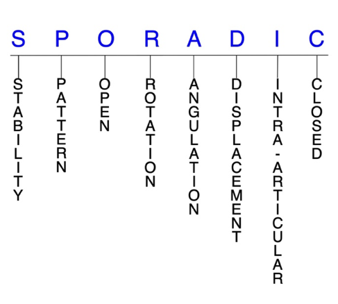
In addition, anatomically named fractures are often also identified by specific eponyms or other special features.

For the Radial Head Fractures, the historical and specifically named fractures include:

Essex-Lopresti injury (ELI)

Terrible Triad Injury (TTI) of the elbow

By selecting the name (diagnosis), you will be linked to the introduction section of this Diagnostic Guide dedicated to the selected fracture eponym.


Fractures of the radial head are the most common of all injuries to the elbow, accounting for nearly one-third of all elbow fractures. These fractures result from direct or indirect trauma to the elbow, most frequently from a fall onto an outstretched hand (FOOSH) with the elbow partially flexed and pronated. Although isolated radial head fractures are possible, they are more likely to occur with associated injuries, particularly elbow dislocations, capitellar fractures, and medial collateral ligament (MCL) tears. Nonoperative treatment is generally indicated for most isolated, stable, and nondisplaced-to-minimally displaced fractures, while surgery is typically required for unstable fractures, displaced fractures, and those that fail conservative management.1-5

Definitions

  • A radial head fracture is a disruption of the mechanical integrity of the radial head.
  • A radial head fracture produces a discontinuity in the radial head contours that can be complete or incomplete.
  • A radial head fracture is caused by a direct force that exceeds the breaking point of the bone.

Hand Surgery Resource’s Fracture Description and Characterization Acronym

SPORADIC

S – Stability; P – Pattern; O – Open; R – Rotation; A – Angulation; D – Displacement; I – Intra-articular; C – Closed


S - Stability (stable or unstable)

  • Universally accepted definitions of clinical fracture stability are not well defined in the literature.6-8
  • Stable: fracture fragment pattern is generally nondisplaced or minimally displaced. It does not require reduction, and the fracture fragments’ alignment is maintained by with simple splinting or casting. However, most definitions define a stable fracture as one that will maintain anatomical alignment after a simple closed reduction and splinting. Some authors add that stable fractures remain aligned, even when adjacent joints are put to a partial range of motion (ROM).
  • Unstable: will not remain anatomically or nearly anatomically aligned after a successful closed reduction and immobilization. Typical unstable radial head fractures have significant deformity with comminution, displacement, angulation, and/or shortening.
  • Unstable radial head fractures—such as fracture-dislocations—require restoration of radiocapitellar contact through radial head repair or reconstruction.1

P - Pattern1,9

  • Type I: nondisplaced or minimally displaced (< 2 mm) fracture of the radial head or neck
  • Type II: displaced (>2 mm) and angulated fracture of the radial head or neck
  • Type III: severely comminuted fracture involving the entire radial head and neck
  • Type IV: radial head fracture with associated elbow dislocation

O - Open

  • Open: a wound connects the external environment to the fracture site. The wound provides a pathway for bacteria to reach and infect the fracture site. As a result, there is always a risk for chronic osteomyelitis. Therefore, open fractures of the radial head require antibiotics with surgical irrigation and wound debridement.6,10,11

R - Rotation

  • Radial head fracture deformity can be caused by proximal rotation of the fracture fragment in relation to the distal fracture fragment.
  • Degree of malrotation of the fracture fragments can be used to describe the fracture deformity.

A - Angulation (fracture fragments in relationship to one another)

  • Angulation is measured in degrees after identifying the direction of the apex of the angulation.
  • Straight: no angulatory deformity
  • Angulated: bent at the fracture site
  • Type II radial head fractures are angulated.12

D - Displacement (Contour)

  • Displaced: disrupted cortical contours
  • Nondisplaced: ≥1 fracture lines defining one or several fracture fragments; however, the external cortical contours are not significantly disrupted

I - Intra-articular involvement

  • Intra-articular fractures are those that enter a joint with ≥1 of their fracture lines.
  • Radial head fractures can have fragment involvement at the proximal radioulnar joint (PRUJ) or radiocapitellar joint.
  • If a fracture line enters a joint but does not displace the articular surface of the joint, then it is unlikely that this fracture will predispose to post-traumatic osteoarthritis. If the articular surface is separated or there is a step-off in the articular surface, then the congruity of the joint will be compromised, and the risk of post-traumatic osteoarthritis increases significantly.

C - Closed

  • Closed: no associated wounds; the external environment has no connection to the fracture site or any of the fracture fragments.4-6

Radial head fractures: named fractures, fractures with eponyms and other special fractures

Essex-Lopresti Injury

  • The Essex-Lopresti injury (ELI) consists of a comminuted radial head fracture combined with a disruption of the distal radioulnar joint (DRUJ) and a tear of the interosseous membrane (IOM).13
    • A fracture-dislocation, dislocation, or triangular fibrocartilage complex tear may also occur in this injury pattern.14
  • These injuries typically occur when a high-energy load is applied axially to the forearm, most often from a FOOSH.13
  • ELIs are uncommon, with one study finding that they were present in only 4% of all radial head fractures.13 Many cases — up to two-thirds — are also unrecognized, usually due to a physician’s failure to perform a complete and thorough examination of the wrist and forearm.14

Imaging

  • Radiology studies - X-ray
    • Anteroposterior (AP), lateral, and oblique views of both the elbow and wrist should be performed, while comparison views of the contralateral wrist or elbow may also be helpful.14
  • Magnetic resonance imaging - MRI without contrast
    • May be performed due to its highest sensitivity for diagnosing IOM tears.4

Treatment14

  • Surgery is typically required for these injuries, and surgical options include the following:
    • Open reduction and internal fixation (ORIF)
    • Radial head replacement
    • Radial head excision
    • DRUJ reduction
    • Wrist arthroscopy, debridement, and a joint leveling procedure (eg, ulnar shortening osteotomy)
    • Interosseous ligament reconstruction

Complications

  • Stiffness
  • Infection
  • Reduced elbow ROM
  • Residual pain
  • Elbow instability
  • Nonunion
  • Failure of fixation

Outcomes

  • ELIs are often associated with unsatisfactory functional outcomes, and outcomes are typically worse with greater treatment delays.4,13
  • Management of comminuted radial head fractures remains controversial, as fixation of more than three parts may lead to early failure of fixation, nonunion, and limited forearm rotation.13

Terrible Triad Injury of the Elbow

  • The terrible triad injury (TTI) of the elbow involves a radial head fracture combined with an elbow dislocation and ulnar coronoid process fracture. This injury has earned its nickname because it is very challenging to treat successfully.13,15
  • TTIs are typically caused by a high-energy FOOSH.13
  • These injuries are relatively rare, accounting for about 8–11% of all elbow dislocations and 3–10% of all radial head fractures.13

Imaging

  • Radiology studies - X-ray
  • Magnetic resonance imaging - MRI without contrast
  • Radiology studies - Computerized tomography (CT) scanning

Treatment

  • TTIs nearly always require surgery, and most experts recommend early surgical intervention to improve the odds of a successful outcome.4,15
  • Surgery typically includes radiocapitellar joint restoration and reattachment of the lateral collateral ligament (LCL), while coronoid fracture fixation and MCL repair may also be performed in some cases.13
    • Several approaches can be used to address coronoid fractures in TTIs, and since there is no consensus on which approach is best, decisions should be made based on fragment size and fracture location.
    • For comminuted radial head fractures, either fixation or artificial replacement can be performed.

Complications

  • Elbow stiffness
  • Heterotopic ossification
  • Elbow joint pain
  • Arthritis
  • Instability

Outcomes

  • TTIs are generally associated with poor long-term outcomes, as complications like stiffness, pain, arthritis, and joint instability are common and can delay recovery.4
  • One study found that radial head replacement allowed for elbow stability to be achieved with comparable overall outcomes to fixation, but the study lacked data on long-term outcomes.13

Related Anatomy3,16-18

  • The elbow is a hinge-type synovial joint comprised of the radius, ulna, and humerus, and formed by three articulations: the ulnohumeral joint, radiocapitellar joint, and PRUJ.
  • The radiocapitellar joint is the articulation of the radial head with the capitellum of the humerus. It is essential to elbow longitudinal and valgus stability and has an integral relationship with the LCL.
    • The radius consists of a rectangular epiphysis at its distal end, a long shaft, and a radial neck and head at its proximal end. The radial head is important because it influences all three elbow articulations. The stability of the radiocapitellar joint is based on the opposite congruity of the convex capitellum with the concave radial head. Articular cartilage covers this concave surface and at least an arc of ~280° around the rim of the radial head.
  • The PRUJ is formed by the articulation of the radial head with the lesser sigmoid notch of the proximal ulna and is stabilized by the annular ligament.
  • The key ligaments of the elbow include the LCL (which extends from the lateral epicondyle and blends with the annular ligament of the radius), the MCL (which originates from the medial epicondyle and attaches to the coronoid process and olecranon of the ulna), and the annular ligament (which encircles the radial head and stabilizes the PRUJ and radiocapitellar joint).
  • The key tendons of the elbow include the tendons associated with the biceps, triceps, and extensor carpi radialis longus (ECRL) muscles as well as the common extensor tendon (the shared origin of the extensor carpi radialis brevis (ECRB), extensor digitorum communis (EDC), extensor digiti minimi (EDM) and extensor carpi ulnaris (ECU)), and the common flexor tendon (the shared origin of the pronator teres, flexor carpi radialis (FCR), palmaris longus, flexor digitorum superficialis (FDS), and flexor carpi ulnaris (FCU)).
  • The radius and ulna are also connected by a sheet of thick fibrous tissue called the IOM.

Incidence

  • Radial head fractures are the most common elbow injuries, accounting for ~30% of elbow fractures and 1–4% of all fractures in adults.5,18
    • Radial head and neck fractures are also responsible for about 0.2% of all visits to the emergency department.3
  • The reported incidence of radial head fractures is between 2.5–2.8 per 10,000 individuals each year.3,4
  • The average age of patients who suffer radial head fractures is 45 years.3
  • Associated injuries are detected in about one-third of radial head fractures, with elbow dislocation occurring in up to 14% and ulnar fracture occurring in up to 12% of cases.3,4
ICD-10 Codes
  • RADIAL HEAD FRACTURE - ADULT

    Diagnostic Guide Name

    RADIAL HEAD FRACTURE - ADULT

    ICD 10 Diagnosis, Single Code, Left Code, Right Code and Bilateral Code

    DIAGNOSISSINGLE CODE ONLYLEFTRIGHTBILATERAL (If Available)
    RADIAL HEAD FRACTURE - ADULT    
    - DISPLACED S52.122_S52.121_ 
    - NONDISPLACED S52.125_S52.124_ 

    Instructions (ICD 10 CM 2020, U.S. Version)

    THE APPROPRIATE SEVENTH CHARACTER IS TO BE ADDED TO EACH CODE FROM CATEGORY S52
     Closed FracturesOpen Type I or II or OtherOpen Type IIIA, IIIB, or IIIC
    Initial EncounterABC
    Subsequent Routine HealingDEF
    Subsequent Delayed HealingGHJ
    Subsequent NonunionKMN
    Subsequent MalunionPQR
    SequelaSSS

    ICD-10 Reference

    Reproduced from the International statistical classification of diseases and related health problems, 10th revision, Fifth edition, 2016. Geneva, World Health Organization, 2016 https://apps.who.int/iris/handle/10665/246208

Clinical Presentation Photos and Related Diagrams
Adult Left Radial Head Fracture
  • Adult left radial head fracture with displaced fragment (arrow) and comminution
    Adult left radial head fracture with displaced fragment (arrow) and comminution
Symptoms
History of trauma
Fracture pain
Fracture deformity
Swelling, ecchymosis and/or tenderness
Abrasion
Typical History

The typical patient is a 43-year-old man who injured himself while playing softball. During a game, he attempted to dive over the second baseman—who was crouching—into second base and landed with both hands outstretched and his right elbow partially flexed and pronated. The force of the impact led to a radial head fracture and a posterior elbow dislocation, and the man experienced immediate pain, swelling, and deformity after the injury.

Positive Tests, Exams or Signs
Work-up Options
Images (X-Ray, MRI, etc.)
Adult left Radial Head Fracture
  • Adult left radial head fracture with displaced fragment (arrow) and comminution
    Adult left radial head fracture with displaced fragment (arrow) and comminution
  • Adult left radial head fracture CT with displaced fragment and comminution
    Adult left radial head fracture CT with displaced fragment and comminution
Treatment Options
Treatment Goals
  • When treating closed radial head fractures, the treating surgeon has 4 basic goals:6,11
    1. An elbow with a normal appearance. The X-ray may not need to be perfect, but the elbow should have no obvious deformity (ie, the elbow looks normal!)
    2. Avoid elbow stiffness by maintaining a normal functional ROM (ie, the elbow works!)
    3. The elbow is not painful (ie, the elbow does not hurt!)
    4. Congruent joint surface with none-to-minimal joint surface irregularities (ie, the elbow does not develop early post-traumatic arthritis!)
  • One additional goal is mandatory for open fractures:
    1. Fracture care should minimize the risk for infection and osteomyelitis.
Conservative
  • Nonoperative treatment is generally indicated for the following radial head fractures: 4,5,19
    • Most isolated fractures
    • Nondisplaced and stable fractures
    • Minimally displaced (<2 mm) partial articular fractures
    • Fractures with joint surface <30% or angulation <30°
  • Type I radial head fractures are typically managed conservatively with a supportive sling followed by early mobilization. Another approach is to splint the elbow at 60° flexion for 1–2 weeks, then allowing unrestricted movement after this period.
  • Some type II radial head fractures may also be treated conservatively, while others may require surgery.
Operative
  • Surgical treatment of radial head fractures must always be an individualized therapeutic decision.1,5 However, surgical radial head fracture care is most frequently recommended when:
    1. Closed reduction fails or the simple splint or cast immobilization does not maintain the reduction. For these irreducible or unstable fractures, operative treatment is recommended to achieve the 4 treatment goals of fracture care.
    2. There is a significantly displaced radial head fracture involving the PRUJ or radiocapitellar joint.
    3. Open radial head fractures. These injuries require surgical care in the form of irrigation and debridement to prevent chronic infection.
  • ORIF
    • Preferred surgical approach for most radial head fractures.
    • Fixation devices include:
      • K-wires
      • Reconstruction plates
      • Mini-condylar plates
      • Mini-fragment screws
      • Herbert mini screws
      • Radial head plate
    • Radial head excision
      • Indicated for some low-demand older adults with complex fractures but no associated fractures that compromise stability.
    • Fragment excision
      • Indicated in when bone fragments are obstructing a joint and the fragments are too small for osteosynthesis
    • Osteosynthesis
      • Primarily indicated for isolated fractures that cause a joint blockage and type II fractures with step-off >2 mm, joint fragment over 30%, and/or radial neck fractures with angulation >30°.
      • Low profile plates and screw implants or cannulated screws are typically recommended.
    • Arthroplasty
      • Primarily indicated for comminuted fractures comprising greater than 25% of the radial head that are not amenable to osteosynthesis (typically those with > 3 fragments)
Treatment Photos and Diagrams
  • Adult left radial head fracture with displaced fragment and comminution treated by radial head replacement
    Adult left radial head fracture with displaced fragment and comminution treated by radial head replacement
  • The care and precautions related to immobilization devices for the radial head fracture must be carefully reviewed with the patient. Patients should be educated regarding care and precautions. Patients should know that pain, especially increasing pain, numbness, tingling, skin irritation, splint loosening, or excessive tightness are red flags and should be reported to the surgeon or his team.
  • Pain should be managed with properly fitting splints and casts, reassurance, elevation, ice in the initial post-fracture period, and mild pain medications. Patients should be encouraged to discontinue pain medication as soon as possible. Opioid use should be kept to a minimum.
  • Joints that are splinted for closed stable fractures are usually immobilized.
  • Fractures that require internal fixation can be mobilized after 4 weeks.
  • Rehabilitation is primarily dependent on the presence of associated injuries and the stability of the repair, with a more detailed rehabilitation program being needed for complex injuries. Most experts recommend early active ROM once the repaired structures are secure. A collar-and-cuff sling is used for the first 2 weeks following surgery, and a static progressive extension splint may be needed at 6 weeks if recovery of extension is slow. Passive stretching can then begin at 6 weeks, and strengthening exercises may be introduced 8–12 weeks after surgery, so long as the osseous and ligamentous injuries have healed.2
Complications
  • Stiffness
  • Malunion
  • Loss of elbow ROM
  • Heterotopic ossification
  • Hardware failure
  • Nonunion
  • Post-traumatic arthritis
  • Avascular necrosis
Outcomes
  • Type I radial head fractures treated conservatively usually heal well with minimal to no complications.4
    • Some studies have shown that conservative treatment involving early active ROM can even lead to positive long-term outcomes for two-part radial head fractures with 2–5 mm of displacement, no block to elbow or forearm motion, and a stable elbow.19
  • One study found that 81% of patients experienced good-to-excellent outcomes at a mean of 22 years after ORIF was performed for isolated, stable, displaced, partial articular radial head fractures, but these outcomes were not superior to another series in which conservative treatment was used to treat similar injuries.19
Key Educational Points
  • Surgeons should be aware that comminuted radial head fractures are likely to have associated bone and/or ligamentous injuries and must treat these associated injuries appropriately to prevent persistent instability. Treatment and prognosis is closely related to what other injuries are identified when the patient initially presents.1,5
  • It is also imperative for surgeons to identify displaced unstable fractures and determine whether reduction and fixation is possible or if arthroplasty is necessary to prevent long-term complications.18
  • Radial head excision was once frequently utilized because the radial head was considered insignificant and believed to have little influence on elbow anatomy and biomechanics, but long-term outcomes associated with this procedure have been poor, and the standard of care has since shifted away from this practice.5
  • Joint exam - It is important to evaluate joint range of motion and stability.  If pain limits assessment, an intra-articular injection with local anesthetic may be given.
  • AP, oblique, and lateral X-ray views are a good starting point and are effective for identifying radial head fractures; however, other associated fractures can be more subtle and difficult to detect with plain radiography. Thus, advanced imaging may be needed if concomitant injury is suspected.2,5,18 Plain radiographs should be particularly scrutinized for fractures of the capitellum, trochlea, or coronoid.2 Radiographs of the forearm and wrist may also be needed if associated injuries seem likely.4 
  • Computerized tomography (CT) scanning may be required if a more complex injury is suspected or the fracture is comminuted.4 Particularly helpful for quantifying displacement and allowing for more accurate assessments of the reparability of fracture fragments.2
  • MRI without contrast is rarely necessary, but when used, can provide additional information about associated soft tissue injuries of the collateral ligaments and the IOM.2
References

Cited Articles

  1. Tejwani, NC and Mehta, H. Fractures of the radial head and neck: current concepts in management. J Am Acad Orthop Surg 2007;15(7):380-7. PMID: 17602027
  2. Pike, JM, Athwal, GS, Faber, KJ, et al. Radial head fractures--an update. J Hand Surg Am 2009;34(3):557-65. PMID: 19258159
  3. van Riet, RP, van den Bekerom, MPJ, Tongel, AV, et al. Radial head fractures. Shoulder & Elbow 2020;12(3):212–223. PMID: 32565923
  4. Khawar, H, Craxford, S and Ollivere, B. Radial head fractures. Br J Hosp Med (Lond) 2020;81(4):1-6. PMID: 32339017
  5. Patiño, JM and Saenz, VP. Radial Head Fractures. In: StatPearls.Treasure Island (FL): 2021. PMID: 28846251
  6. Cheah, AE and Yao, J. Hand Fractures: Indications, the Tried and True and New Innovations. J Hand Surg Am 2016;41(6):712-22. PMID: 27113910
  7. Nesbitt, KS, Failla, JM and Les, C. Assessment of instability factors in adult distal radius fractures. J Hand Surg Am 2004;29(6):1128-38. PMID: 15576227
  8. Walenkamp, MM, Vos, LM, Strackee, SD, et al. The Unstable Distal Radius Fracture-How Do We Define It? A Systematic Review. J Wrist Surg 2015;4(4):307-16. PMID: 26649263
  9. Hotchkiss, RN. Displaced Fractures of the Radial Head: Internal Fixation or Excision? J Am Acad Orthop Surg 1997;5(1):1-10. PMID: 10797202
  10. Ketonis, C, Dwyer, J and Ilyas, AM. Timing of Debridement and Infection Rates in Open Fractures of the Hand: A Systematic Review. Hand (N Y) 2017;12(2):119-126. PMID: 28344521
  11. Meals, C and Meals, R. Hand fractures: a review of current treatment strategies. J Hand Surg Am 2013;38(5):1021-31. PMID: 23618458
  12. Iannuzzi, NP and Leopold, SS. In brief: the Mason classification of radial head fractures. Clin Orthop Relat Res 2012;470(6):1799-802. PMID: 22426693
  13. Shinohara, I, Takahara, S, Uefuji, A, et al. Floating forearm associated with terrible triad injury and Essex-Lopresti injury: a case report and literature review. JSES Int 2021;5(2):320-327. PMID: 33681857
  14. Adams, JE and Osterman, AL. The Essex-Lopresti Injury:: Evaluation and Treatment Considerations. Hand Clin 2020;36(4):463-468. PMID: 33040958
  15. He, X, Fen, Q, Yang, J, et al. Risk Factors of Elbow Stiffness After Open Reduction and Internal Fixation of the Terrible Triad of the Elbow Joint. Orthop Surg 2021;13(2):530-536. PMID: 33619861
  16. Fernandez, DL and Jupiter, JB. Fractures of the Distal Radius: A Practical Approch to Management. Second ed. New York: Springer Science+Business Media New York; 2002.
  17. Zumstein, MA, Hasan, AP, McGuire, DT, et al. Distal radius attachments of the radiocarpal ligaments: an anatomical study. J Wrist Surg 2013;2(4):346-50. PMID: 24436840
  18. Burkhart, KJ, Wegmann, K, Muller, LP, et al. Fractures of the Radial Head. Hand Clin 2015;31(4):533-46. PMID: 26498543
  19. Ruchelsman, DE, Christoforou, D and Jupiter, JB. Fractures of the radial head and neck. J Bone Joint Surg Am 2013;95(5):469-78. PMID: 23467871
Subscribe to RADIAL HEAD FRACTURE ADULT