Introduction

Fracture Nomenclature for Humeral Shaft fractures

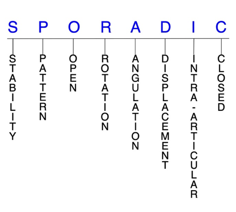
Hand Surgery Resource’s Diagnostic Guides describe fractures by the anatomical name of the fractured bone and then characterize the fracture by the Acronym:
In addition, anatomically named fractures are often also identified by specific eponyms or other special features.

For the Humeral Shaft Fractures, the historical and specifically named fractures include:

Holstein-Lewis fracture

By selecting the name (diagnosis), you will be linked to the introduction section of this Diagnostic Guide dedicated to the selected fracture eponym.


Humeral shaft fractures are common injuries that most frequently occur in men aged 21–30 and women aged 60–80. In younger patients, these injuries usually occur due to a direct blow to the arm (e.g., car accidents, sports injuries), while in older patients, the mechanism of injury is a fall on an outstretched hand in which the humerus takes the brunt of the impact. Many patients with humeral shaft fractures have concomitant radial nerve injury due to the proximity of the nerve to the bone. Most humeral shaft fractures are amenable to conservative treatment consisting of immobilization and/or functional bracing, and positive outcomes are likely with this approach. Surgery may be indicated in patients with extensive concomitant injury and if conservative treatment fails.1-3

Definitions

  • A humeral shaft fracture is a disruption of the mechanical integrity of the humeral shaft.
  • A humeral shaft fracture produces a discontinuity in the humeral shaft contours that can be complete or incomplete.
  • A humeral shaft fracture is caused by a direct force that exceeds the breaking point of the bone.

Hand Surgery Resource’s Fracture Description and Characterization Acronym

SPORADIC

S – Stability; P – Pattern; O – Open; R – Rotation; A – Angulation; D – Displacement; I – Intra-articular; C – Closed


S - Stability (stable or unstable)

  • Universally accepted definitions of clinical fracture stability are not well defined in the literature.4-6
  • Stable: fracture fragment pattern is generally nondisplaced or minimally displaced. It does not require reduction, and the fracture fragments’ alignment is maintained by with simple immobilization. However, most definitions define a stable fracture as one that will maintain anatomical alignment after a simple closed reduction and immobilization. Some authors add that stable fractures remain aligned, even when adjacent joints are put to a partial range of motion (ROM).
    • Most humeral shaft fractures are unstable, but conservative management is still considered the gold standard in most cases.1
  • Unstable: will not remain anatomically or nearly anatomically aligned after a successful closed reduction and immobilization. Typical unstable humeral shaft fractures have significant deformity with comminution, displacement, angulation, and/or shortening.

P - Pattern1,3

  • Type A: simple fractures
    • Type A1: spiral
    • Type A2: oblique
    • Type A3: transverse
  • Type B: wedge fractures
    • Type B1: spiral (intact)
    • Type B2: bending (intact)
    • Type B3: fragmented
  • Type C: complex/multifragmentary fractures
    • Type C1: spiral
    • Type C2: segmental
    • Type C3: irregular

O - Open

  • Open: a wound connects the external environment to the fracture site. The wound provides a pathway for bacteria to reach and infect the fracture site. As a result, there is always a risk for chronic osteomyelitis. Therefore, open fractures of the humeral shaft require antibiotics with surgical irrigation and wound debridement.4,7,8

R - Rotation

  • Humeral shaft fracture deformity can be caused rotation of the fracture proximal fragment in relation to the distal fracture fragment.
  • Degree of malrotation of the fracture fragments can be used to describe the fracture deformity.

A - Angulation (fracture fragments in relationship to one another)

  • Angulation is measured in degrees after identifying the direction of the apex of the angulation.
  • Straight: no angular deformity
  • Angulated: bent at the fracture site

D - Displacement (Contour)

  • Displaced: disrupted cortical contours
  • Nondisplaced: ≥1 fracture lines defining one or several fracture fragments; however, the external cortical contours are not significantly disrupted
  • Holstein-Lewis fractures involve displacement of the distal fracture fragment and a simple spiral fracture pattern.2

I - Intra-articular involvement

  • Intra-articular fractures are those that enter a joint with ≥1 of their fracture lines.
  • Humeral shaft fractures can extend to have fragment involvement at the glenohumeral, ulnohumeral, or radiocapitellar joints.
  • If a fracture line enters a joint but does not displace the articular surface of the joint, then it is unlikely that this fracture will predispose to post-traumatic osteoarthritis. If the articular surface is separated or there is a step-off in the articular surface, then the congruity of the joint will be compromised, and the risk of post-traumatic osteoarthritis increases significantly.

C - Closed

  • Closed: no associated wounds; the external environment has no connection to the fracture site or any of the fracture fragments.4-6

Humeral shaft fractures: named fractures, fractures with eponyms and other special fractures

Holstein-Lewis fracture

  • The Holstein-Lewis fracture pattern is a simple spiral fracture of the distal humeral shaft in which the distal fragment is displaced, and the proximal end is deviated towards the radial side.2,9
  • Concomitant radial nerve injury is common in Holstein-Lewis fractures because the distal fragment can potentially lacerate or entrap the radial nerve after it is displaced.2,9
  • Holstein-Lewis fractures account for approximately 7.5% of all humeral shaft fractures and are more common in younger patients following high-energy trauma.2

Imaging

  • Radiology studies - X-ray
    • Radiographs are usually sufficient to diagnose these injuries.10

Treatment

  • Although it was once believed that surgery was necessary for Holstein-Lewis fractures, most of these injuries are now treated nonsurgically. Conservative treatment includes immobilization using a coaptation splint with a collar and cuff or sling and swathe.10

Complications

  • Radial nerve palsy
  • Nonunion
  • Stiffness

Outcomes

  • According to one study, long-term outcomes were comparable between patients with Holstein-Lewis fractures who were treated conservatively and surgically.9

Related Anatomy2,3,11

  • The humerus is a long bone that can be divided into a proximal end, a long shaft, and a distal end.
    • The proximal end consists of an anatomic neck, the humeral head, the surgical neck, and the greater and lesser tuberosities at its proximal end. The humeral head humerus articulates with the glenoid fossa of the scapula to form the glenohumeral joint.
    • The humeral shaft extends distally from the proximal border of the pectoralis major insertion to the supracondylar ridge. It is nearly cylindrical in its proximal half and then becomes flattened and triangular towards its distal end. It has 3 major surfaces: the anterolateral, anteromedial, and posterior surfaces.
    • At the distal end, the medial condyle articulates with the ulna to form the ulnohumeral joint, and the capitellum articulates with the radial head to form the radiocapitellar joint.
  • The humeral shaft serves as an insertion site for the pectoralis major, deltoid, and coracobrachialis tendons, and is the origin site for the brachialis, triceps, and brachioradialis tendons.
  • Two important regions of the humeral shaft are the deltoid tuberosity and the radial groove.
    • The deltoid tuberosity is an elevation near the middle of the anterolateral surface, which is the insertion point of the deltoid tendon.
    • The radial groove or sulcus starts distal to the attachment of the lateral head of triceps on the posterior surface and runs distal and lateral toward the anterolateral surface. The radial nerve and profunda artery both pass within this groove.
  • The radial nerve is the major nerve of the humeral shaft, which is located 14 cm proximal to the lateral epicondyle and 20 cm proximal to the medical epicondyle. Its proximity to the osseous anatomy of the humerus explains why the radial nerve is at such a high risk for injury in humeral shaft fractures.

Incidence

  • Humeral shaft fractures account for about 1–3% of all fractures.12
  • The incidence of humeral shaft fractures is approximately 13 per 100,000 persons per year, and about 66,000 humeral shaft fractures occur annually in the U.S.2,3
  • The bimodal peaks for humeral shaft fractures are in men aged 21–30 years and women aged 60–80 years.1
  • About 30% of humeral shaft fractures occur in the proximal third, 60% in the middle third, and 10% in the distal third of the bone.1

ICD-10 Codes
  • HUMERAL SHAFT FRACTURE - ADULT

    Diagnostic Guide Name

    HUMERAL SHAFT FRACTURE - ADULT

    ICD 10 Diagnosis, Single Code, Left Code, Right Code and Bilateral Code

    DIAGNOSISSINGLE CODE ONLYLEFTRIGHTBILATERAL (If Available)
    HUMERAL SHAFT FRACTURE - ADULT    
    TORUS FRACTURE LOWER HUMERUS    
    - DISPLACED S42.482_S42.481_ 
    - NONDISPLACED S42.435_S42.434_ 
    GREENSTICK S42.312_S42.311_ 
    TRANSVERSE    
    - DISPLACED S42.322_S42.321_ 
    - NONDISPLACED S42.325_S42.324_ 
    OBLIQUE    
    - DISPLACED S42.331_S42.332_ 
    - NONDISPLACED S42.335_S42.334_ 
    SPIRAL    
    - DISPLACED S42.432_S42.431_ 
    - NONDISPLACED S42.345_S42.344_ 
    COMMINUTED    
    - DISPLACED S42.352_S42.351_ 
    - NONDISPLACED S42.355_S42.354_ 
    SEGMENTAL    
    - DISPLACED S42.362_S42.361_ 
    - NONDISPLACED S42.365_S42.364_ 

    Instructions (ICD 10 CM 2020, U.S. Version)

    THE APPROPRIATE SEVENTH CHARACTER IS TO BE ADDED TO EACH CODE FROM CATEGORY S42
     Closed FracturesOpen Type I or II or OtherOpen Type IIIA, IIIB, or IIIC
    Initial EncounterABC
    Subsequent Routine HealingDEF
    Subsequent Delayed HealingGHJ
    Subsequent NonunionKMN
    Subsequent MalunionPQR
    SequelaSSS

    ICD-10 Reference

    Reproduced from the International statistical classification of diseases and related health problems, 10th revision, Fifth edition, 2016. Geneva, World Health Organization, 2016 https://apps.who.int/iris/handle/10665/246208

Clinical Presentation Photos and Related Diagrams
Humeral Shaft fracture
  • Closed right transverse mid-shaft minimally displaced humerus fracture
    Closed right transverse mid-shaft minimally displaced humerus fracture
Symptoms
History of trauma
Fracture pain
Fracture deformity
Swelling, ecchymosis & tenderness
Abrasion
Typical History

A typical patient is a 22-year-old woman who was injured in a car accident. The woman was riding in the passenger seat of her friend’s car and traveling at a fast speed (>80 MPH) on the highway when her friend failed to notice that traffic had come to a complete stop ahead. The driver suddenly slammed on the brakes but swerved to the right after realizing the car would not stop in time. As a result, their car collided with another car in the right lane, and this collision resulted in a fracture the humeral shaft of the patient’s right arm due to the impact of the car door. Both the driver and passenger were taken to the emergency department, where the passenger was treated for a Type C1 humeral shaft fracture.

Positive Tests, Exams or Signs
Work-up Options
Images (X-Ray, MRI, etc.)
Humerus Shaft Fracture
Treatment Options
Treatment Goals
  • When treating closed humeral shaft fractures, the treating surgeon has 4 basic goals:4,8
    1. A humerus with a normal appearance. The X-ray may not need to be perfect, but the humerus should have no obvious deformity (i.e., the humerus looks normal!)
    2. Avoid shoulder and elbow stiffness by maintaining a normal functional ROM (i.e., the shoulder and elbow work!)
    3. The humerus is not painful (i.e., the humerus does not hurt!)
    4. Congruent joint surface with none-to-minimal joint surface irregularities (i.e., the shoulder and elbow do not develop early post-traumatic arthritis!)
  • One additional goal is mandatory for open fractures:
    1. Fracture care should minimize the risk for infection and osteomyelitis.
Conservative
  • Most humeral shaft fractures can be treated nonsurgically. Aside from the typical indications for surgical treatment discussed below, some surgeons prefer to also treat fractures of the distal third of the humeral shaft surgically; however, evidence is lacking to support surgery as superior for these fractures, meaning surgeons should make this determination on a case-by-case basis1,2.
  • Conservative treatment for humeral shaft fractures involves reduction through immobilization with a splint, followed by functional bracing.
  • Before splinting, the clinician should address any overlying breaks in the skin (eg, lacerations, abrasions, or skin tears). Splinting options for a humeral shaft fracture include a coaptation splint with a sling, a hanging arm cast, a long arm splint, or a shoulder immobilizer.
  • Since immobilization of the humeral shaft is difficult, both the elbow and shoulder should also be immobilized.
  • Immobilization should be kept in place for 1–2 weeks. If the acute symptoms have subsided and soft tissue conditions are permissive at the follow-up examination, a functional, adjustable brace is then applied and worn until osseous union, which is usually 10–12 weeks.
Operative
  • Surgical treatment of humeral shaft fractures must always be an individualized therapeutic decision.1-3 However, surgical humeral shaft fracture care is most frequently recommended when:
    1. Closed reduction fails or the simple immobilization does not maintain the reduction. For these irreducible or unstable fractures, operative treatment is recommended to achieve the 4 treatment goals of fracture care.
    2. There is a significantly displaced humeral shaft fracture involving the glenohumeral, ulnohumeral, and/or radiocapitellar joint.
    3. Open humeral shaft fractures. These injuries require surgical care in the form of irrigation and debridement to prevent chronic infection.
  • Additional surgical indications include the presence of vascular injury or nerve injury, intra-articular fractures, floating elbow, high-velocity gunshot wounds, comminuted fractures, and the inability to tolerate a splint or brace for a prolonged period.
  • Open reduction and internal fixation (ORIF)
    • Often regarded as the preferred technique for humeral shaft fractures requiring surgery, but especially indicated for open fractures and those with concomitant vascular injury requiring repair, brachial plexus injury, floating elbow, or compartment syndrome.
      • Multiple surgical exposures can be selected, many of which can also be extended to address concomitant fractures around the shoulder and elbow.
    • Various plate options are available, with large fragment plates—both broad and narrow—being used most frequently, but small fragment plates and mini-fragment plates also serving a role at times.
  • External fixation
    • May be considered for patient with polytrauma, bilateral fractures, or severe fracture comminution or soft tissue injury, but used less frequently due to the advancements of other surgical techniques.
  • Minimally invasive percutaneous osteosynthesis
    • Emerging treatment option for highly comminuted humeral shaft fractures, since it limits the amount of dissection needed compared to conventional plating.
    • Typically performed through an anterior approach, which protects the radial nerve.
  • Intramedullary nailing
    • Generally indicated for pathologic fractures, segmental fractures, and patients with severe osteoporotic bone, overlying skin compromise, or polytrauma.
    • Can be performed antegrade or retrograde. However, antegrate nailing is much more commonly performed. Retrograde nailing may sometimes be used for mid-diaphyseal or proximal third fractures, and occasionally distal third fractures.
    • Intramedullary locked nails are used most often because they allow preservation of periosteal blood supply and minimize disruption of fracture biology. Flexible nails, Kirschner wires, and Enders nails are used less frequently.
Treatment Photos and Diagrams
Right Adult Treatment
  • Humerus fracture treated with a coaptation splint.
    Humerus fracture treated with a coaptation splint.
Humerus Fracture Treatment Options
  • Humerus fracture with 100% displacement
    Humerus fracture with 100% displacement
  • Displaced humerus fracture treated with open reduction and internal fixation (ORIF) with plate and screws.
    Displaced humerus fracture treated with open reduction and internal fixation (ORIF) with plate and screws.

Post-treatment Management

  • The care and precautions related to immobilization devices for the humeral shaft fracture must be carefully reviewed with the patient. Patients should be educated regarding care and precautions. Patients should know that pain, especially increasing pain, numbness, tingling, skin irritation, splint loosening, or excessive tightness are red flags and should be reported to the surgeon or his team.
  • Pain should be managed with properly fitting splints and casts, reassurance, elevation, ice in the initial post-fracture period, and mild pain medications. Patients should be encouraged to discontinue pain medication as soon as possible. Opioid use should be kept to a minimum.
  • Joints that are splinted for closed stable fractures are usually immobilized.
  • Fractures that require internal fixation can usually be mobilized either immediately or after 2-4 weeks depending on the mode and adequacy of the internal fixation.
  • After the fracture has healed and the functional brace is removed, patients must undergo an extensive physical therapy program to restore range of motion and muscle mass and strength.1
Complications
  • Nonunion
    • Documented non-union rates following humeral shaft fracture treatment are 1–12%. There is a greater chance for non-union in transverse fractures and in cases with inadequate shoulder mobilization and soft-tissue interposition.1
    • Nonunion rates are also higher in fractures of the proximal third of the humeral shaft compared to fractures of the distal two-thirds.2
  • Radial nerve palsy
  • Angulation
  • Reduced shoulder or elbow ROM
  • Stiffness
  • Aseptic necrosis
  • Avascular necrosis
  • Malunion
  • Heterotopic ossification
  • Hardware failure
  • Infection
  • Post-traumatic arthritis
Outcomes
  • The prognosis for humeral shaft fractures is positive, as conservative treatment has been found to lead to appropriate alignment and union in about 90% of cases.1
    • One study found that in 620 patients with humeral shaft fractures treated with functional bracing, the nonunion rate was only 2% in closed fractures and 6% in open fractures.2
  • Time to union after conservatively treated humeral shaft fractures can vary from 3–5 months.1
  • Research comparing surgical and nonsurgical management of humeral shaft fractures is scarce, but in one study, 44 patients treated with functional bracing (group 1) were compared with 45 patients treated with intramedullary nailing (group 2). Two patients in group 1 and no patients in group 2 experienced nonunion. At the final follow-up, 86% of patients in group 1 and 48% of patients in group 2 had unrestricted shoulder movement.13
  • A systematic review and meta-analysis of humeral shaft fractures reported fewer non-unions but more deep infections in the surgical group compared to the nonoperative group.14
Key Educational Points
  • The biggest controversies surrounding the optimal management humeral shaft fractures involve the types of screws used and the number of cortices and screws needed per fracture segment.3
  • There is currently no failsafe treatment algorithm or strict guidelines for humeral shaft fractures. Thus, clinicians should utilize a share decision-making model with each patient that includes thoughtful discussion with meticulous consideration of patient’s preferences to determine the optimal treatment approach.15
  • Although conservative treatment is currently the gold standard, some studies suggest that the rate of surgical intervention is increasing.14
  • Anteroposterior (AP) and lateral views should be performed in all patients with suspected humeral shaft fractures.  A transthoracic lateral view may be helpful to better visualize any sagittal plane deformity present, while a traction view is not typically necessary but may be helpful for fractures with significant shortening or with a proximal or distal extension.
References

Cited Articles

  1. Bounds, EJ, Frane, N and Kok, SJ. Humeral Shaft Fractures. In: StatPearls. Treasure Island (FL): 2021. PMID: 29292035
  2. Updegrove, GF, Mourad, W and Abboud, JA. Humeral shaft fractures. J Shoulder Elbow Surg 2018;27(4):e87-e97. PMID: 29292035
  3. Carroll, EA, Schweppe, M, Langfitt, M, et al. Management of humeral shaft fractures. J Am Acad Orthop Surg 2012;20(7):423-33. PMID: 22751161
  4. Cheah, AE and Yao, J. Hand Fractures: Indications, the Tried and True and New Innovations. J Hand Surg Am 2016;41(6):712-22. PMID: 27113910
  5. Nesbitt, KS, Failla, JM and Les, C. Assessment of instability factors in adult distal radius fractures. J Hand Surg Am 2004;29(6):1128-38. PMID: 15576227
  6. Walenkamp, MM, Vos, LM, Strackee, SD, et al. The Unstable Distal Radius Fracture-How Do We Define It? A Systematic Review. J Wrist Surg 2015;4(4):307-16. PMID: 26649263
  7. Ketonis, C, Dwyer, J and Ilyas, AM. Timing of Debridement and Infection Rates in Open Fractures of the Hand: A Systematic Review. Hand (N Y) 2017;12(2):119-126. PMID: 28344521
  8. Meals, C and Meals, R. Hand fractures: a review of current treatment strategies. J Hand Surg Am 2013;38(5):1021-31. PMID: 23618458
  9. Ekholm, R, Ponzer, S, Tornkvist, H, et al. The Holstein-Lewis humeral shaft fracture: aspects of radial nerve injury, primary treatment, and outcome. J Orthop Trauma 2008;22(10):693-7. PMID: 18978544
  10. Acuna, J and Hahn, B. Holstein Lewis humerus fracture. Am J Emerg Med 2016;34(2):345 e5-6. PMID: 26166381
  11. Attum, B and Thompson, JH. Humerus Fractures Overview. In: StatPearls. Treasure Island (FL): 2021. PMID: 29489190
  12. Ramo, L, Sumrein, BO, Lepola, V, et al. Effect of Surgery vs Functional Bracing on Functional Outcome Among Patients With Closed Displaced Humeral Shaft Fractures: The FISH Randomized Clinical Trial. JAMA 2020;323(18):1792-1801. PMID: 32396179
  13. Wallny, T, Sagebiel, C, Westerman, K, et al. Comparative results of bracing and interlocking nailing in the treatment of humeral shaft fractures. Int Orthop 1997;21(6):374-9. PMID: 9498146
  14. Lode, I, Nordviste, V, Erichsen, JL, et al. Operative versus nonoperative treatment of humeral shaft fractures: a systematic review and meta-analysis. J Shoulder Elbow Surg 2020;29(12):2495-2504. PMID: 32553853
  15. Driesman, AS, Fisher, N, Karia, R, et al. Fracture Site Mobility at 6 Weeks After Humeral Shaft Fracture Predicts Nonunion Without Surgery. J Orthop Trauma 2017;31(12):657-662. PMID: 28708781
Subscribe to HUMERAL SHAFT FRACTURE ADULT