Introduction

Fracture Nomenclature for Trapezoid Fractures

Hand Surgery Resource’s Diagnostic Guides describe fractures by the anatomical name of the fractured bone and then characterize the fracture by the Acronym:
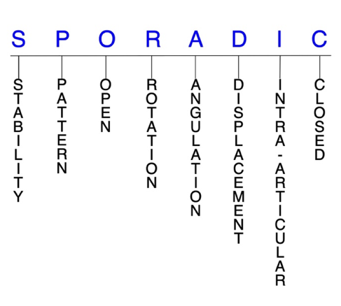
In addition, anatomically named fractures are often also identified by specific eponyms or other special features.

For the Trapezoid Fracture, the historical and specifically named fractures include:

Trapezoid dislocations and fracture-dislocations

By selecting the name (diagnosis), you will be linked to the introduction section of this Diagnostic Guide dedicated to the selected fracture eponym.


Fractures of the carpal bones account for ~6% of fractures overall and up to 18% of all hand fractures. The vast majority (58-89%) occurs in the scaphoid, while fractures of the other 7 carpals are uncommon and only comprise ~1.1% of all fractures. The trapezoid is the least commonly fractured of the carpal bones, with a reported incidence of 0.4-1% of all carpal fractures. This is primarily due to the stability of the trapezoid. Trapezoid fractures rarely occur in isolation and are more frequently associated with other injuries, such as fractures of adjacent carpal bones and disruptions at the carpometacarpal (CMC) and intercarpal joints. Most experts agree that conservative treatment is indicated for nondisplaced and minimally displaced fractures, while surgery is often required for displaced cases, those with articular incongruity, and after nonuion.1-5

Definitions

  • A trapezoid fracture is a disruption of the mechanical integrity of the trapezoid.
  • A trapezoid fracture produces a discontinuity in the trapezoid contours that can be complete or incomplete.
  • A trapezoid fracture is caused by a direct force that exceeds the breaking point of the bone. 

Hand Surgery Resource’s Fracture Description and Characterization Acronym

SPORADIC

S – Stability; P – Pattern; O – Open; R – Rotation; A – Angulation; D – Displacement; I – Intra-articular; C – Closed


S - Stability (stable or unstable)

  • Universally accepted definitions of clinical fracture stability is not well defined in the hand surgery literature.6-8
  • Stable: fracture fragment pattern is generally nondisplaced or minimally displaced. It does not require reduction, and the fracture fragment’s alignment is maintained with simple splinting. However, most definitions define a stable fracture as one that will maintain anatomical alignment after a simple closed reduction and splinting. Some authors add that stable fractures remain aligned, even when adjacent joints are put to a partial range of motion (ROM).
  • Unstable: will not remain anatomically or nearly anatomically aligned after a successful closed reduction and simple splinting. Typically unstable trapezoid fractures have significant deformity with comminution, displacement, angulation, and/or shortening.

P - Pattern

  • Trapezoid dorsal rim
  • Trapezoid body
  • Can be sagittal, coronal, and/or comminuted.3,4
    • When an isolated trapezoid fracture is displaced enough to be see on radiographs, it usually has a characteristic coronal orientation.4
  • The most common mechanism of injury is an axial load through a flexed index metacarpal, which drives the metacarpal proximally and displaces the trapezoid dorsally.
    • Other less common mechanisms include direct trauma and forced flexion-extension through the index MP joint.4,5
    • Due to the rigid stability of the trapezoid within the carpus, high-energy trauma is needed to fracture the bone. This is why associated fractures of surrounding bones are so common.4

O - Open

  • Open: a wound connects the external environment to the fracture site. The wound provides a pathway for bacteria to reach and infect the fracture site. As a result, there is always a risk for chronic osteomyelitis. Therefore, open fractures of the trapezoid require antibiotics with surgical irrigation and wound debridement.6,9,10
  • Open fractures of the trapezoid may require surgical exploration to determine if articular surfaces are involved. After irrigation and debridement, these wounds are generally left open and further treatment is typically delayed until the wound shows no sign of infection.11,12
  • Since high-energy trauma is often needed to cause a trapezoid dislocation, some of these injuries are open.

R - Rotation

  • Trapezoid fracture deformity can be caused by rotation of the distal fragment on the proximal fragment.
  • Degree of malrotation of the fracture fragments can be used to describe the fracture deformity.

A - Angulation (fracture fragments in relationship to one another)

  • Angulation is measured in degrees after identifying the direction of the apex of the angulation.
  • Straight: no angulatory deformity
  • Angulated: bent at the fracture site

D - Displacement (Contour)

  • Displaced: disrupted cortical contours
  • Nondisplaced: fracture line defining one or several fracture fragments; however, the external cortical contours are not significantly disrupted
  • Displaced trapezoid fractures may occur concomitantly with an associated dislocation of the trapezoid or index metacarpal.3

I - Intra-articular involvement

  • Fractures that enter a joint with one or more of their fracture lines.
  • Trapezoid fractures can have fragment involvement at the index CMC joint or at any of its intercarpal joint articulations.
  • If a fracture line enters a joint but does not displace the articular surface of the joint, then it is unlikely that this fracture will predispose to posttraumatic osteoarthritis. If the articular surface is separated or there is a step-off in the articular surface, then the congruity of the joint will be compromised and the risk of posttraumatic osteoarthritis increases significantly.

C - Closed

  • Closed: no associated wounds; the external environment has no connection to the fracture site or any of the fracture fragments.6-8

Trapezoid fractures: named fractures, fractures with eponyms and other special fractures

Trapezoid dislocations and fracture-dislocations

  • Dislocations of the trapezoid are extremely rare injuries that usually occur in conjunction with other hand and wrist injuries. Isolated trapezoid dislocations are even less common.3,13
    • The most common associated injuries are other carpal fractures, trapezium dislocations, and multiple CMC dislocations and fracture-dislocations.13
  • Trapezoid dislocations typically result from high-energy trauma, and possible mechanisms of injury include industrial trauma, motorcycle accidents, and falls from a height. They have also been described as steering wheel injuries, which are caused by powerful axial loading force on the dorsal hand when grasping something strongly.
    • Because many trapezoid dislocations result from serious accidents, they may not be prioritized over other life-threatening injuries. Associated soft-tissue injuries are also common.13,14
  • Dorsal dislocations are far more common than volar dislocations because the volar ligamentous attachments of the trapezoid are stronger than the dorsal ligaments.13
    • Trapezoid fracture-dislocations, in particular, tend to be dorsal and may involve an avulsion of the volar ligaments.3

Imaging

  • Trapezoid dislocations can be missed or ignored because of other life-threatening complications, local swelling, and/or confusing X-rays. A high index of clinical suspicion should be maintained after high-energy injuries to prevent a delayed diagnosis.13,14
  • A straight posteroanterior (PA) radiographic view may be helpful; however, X-rays are often not reliably accurate for diagnosing trapezoid dislocations because of the high prevalence of concomitant injuries.13
  • CT scan
    • Utilized by some surgeons for any high-energy injuries suspected of trapezoid dislocation, because it can more accurately identify injuries when radiographs are inconclusive.14

Treatment

  • There is no consensus regarding the optimal treatment of trapezoid fractures and fracture-dislocations, with different authors reporting a variety of approaches.13
  • Some surgeons insist that anatomical reduction is essential for obtaining a good result, while others have reported that trapezoid resection and limited intercarpal fusion without anatomical reduction is also a viable route.13
  • Closed reduction and percutaneous pinning (CRPP)
    • Can be attempted for some closed trapezoid fracture-dislocations, but the success rate id low.15,16
  • Open reduction and internal fixation (ORIF)
    • Often necessary if CRPP fails.16
  • Limited intercarpal fusion with trapezoid resection
    • May be a useful option for chronic trapezoid dislocations.13
  • Whatever surgical intervention is utilized, soft tissue preservation should be a priority for all surgeons.17

Complications

  • Infection
  • Avascular necrosis

Outcomes

  • Although the management of isolated trapezoid dislocations varies considerably, most patients experience reasonably good outcomes regardless of the treatment used.15
  • Closed reduction has not been successful for treating volar trapezoid dislocations.17

Related Anatomy

  • The trapezoid is an irregular, wedge-shaped bone that consists of a dorsal rim and a body. It is the smallest of the carpals in the distal row and is considered the keystone of the carpal arch, with its dorsal surface being widest and approximately twice the size of its volar surface. The trapezoid articulates with the scaphoid proximally, the capitate ulnarly, the trapezium radially, and the index metacarpal distally.3,18
  • Ligamentous attachments include the trapeziotrapezoid, trapeziocapitate, dorsal and volar CMC, dorsal intercarpal, and triscaphe ligaments. These stout ligaments and the position of the trapezoid keep it protected from most injuries and transfers axial-loaded forces from the index metacarpal base into the scaphoid.3,4,18
  • Tendons associated with the trapezoid include the deep head of flexor pollicis brevis, which originates from its volar surface, and the oblique head of the adductor pollicis, which originates from its distal ulnar volar surface.3

Incidence and Related injuries/conditions

  • Fractures of the carpal bones have been found to account for 8-18% of all hand fractures19,20 and ~6% of fractures overall.21
  • Fractures of the proximal carpals are more common than the distal carpals.
  • Most commonly fractured carpal bone is the scaphoid, representing 58-89% of all carpal fractures.19,20,22,23
  • Other carpal fractures are rare and only account for ~1.1% of all fractures. The triquetrum ranks highest of these bones, while fractures of the remaining carpals are even less common and incidence estimates vary.24-26
  • Trapezoid fractures are the least commonly fractured carpal, having a reported incidence of 0.4-1% of all carpal fractures.26-28
    • Most trapezoid fractures are associated with other carpal bone injuries.
    • In a large case series at a level 1 trauma center over 3 years in, 95% of trapezoid fractures involved concomitant wrist injuries.4
ICD-10 Codes
  • TRAPEZOID FRACTURE

    Diagnostic Guide Name

    TRAPEZOID FRACTURE

    ICD 10 Diagnosis, Single Code, Left Code, Right Code and Bilateral Code

    DIAGNOSISSINGLE CODE ONLYLEFTRIGHTBILATERAL (If Available)
    FRACTURE CARPAL BONE - TRAPEZOID    
    - DISPLACED S62.182_S62.181_ 
    - NONDISPLACED S62.185_S62.184_ 

    Instructions (ICD 10 CM 2020, U.S. Version)

    THE APPROPRIATE SEVENTH CHARACTER IS TO BE ADDED TO EACH CODE FROM CATEGORY S62
     Closed FracturesOpen Type I or II or OtherOpen Type IIIA, IIIB, or IIIC
    Initial EncounterABC
    Subsequent Routine HealingDEF
    Subsequent Delayed HealingGHJ
    Subsequent NonunionKMN
    Subsequent MalunionPQR
    SequelaSSS

    ICD-10 Reference

    Reproduced from the International statistical classification of diseases and related health problems, 10th revision, Fifth edition, 2016. Geneva, World Health Organization, 2016 https://apps.who.int/iris/handle/10665/246208

Clinical Presentation Photos and Related Diagrams
Trapezoid Fracture Diagram
  • Nondisplaced trapezoid body fracture (arrow)
    Nondisplaced trapezoid body fracture (arrow)
Symptoms
History of trauma
Fracture pain, swelling and/or deformity
Impaired wrist ROM
Typical History

A typical patient is a 24-year-old right-handed man who suffered a severe wrist injury in a motor vehicle accident. The man was driving at a fast speed when he failed to notice traffic slowing down ahead of him, and by the time he reacted to slam on the brakes, it was too late to avoid a collision. While still gripping the steering wheel firmly, his car rammed into a stationary car in traffic, and his right hand subsequently endured an extreme axial load from its grip on the steering wheel. This caused fractures of the trapezoid, trapezium, and capitate, as well as several dislocations at these bones’ articulations.

Positive Tests, Exams or Signs
Work-up Options
Treatment Options
Treatment Goals
  • When treating closed trapezoid fractures, the treating surgeon has 5 basic goals:6,10
    1. A trapezoid with a normal appearance. The X-ray may not need to be perfect, but the trapezoid should have no obvious deformity (ie, the trapezoid looks normal!)
    2. Avoid stiffness by maintaining a normal functional ROM (ie, the wrist works!)
    3. The trapezoid is not painful (ie, the trapezoid does not hurt!)
    4. Congruent joint surface with none-to-minimal joint surface irregularities (ie, the joints associated with the trapezoid do not develop early posttraumatic arthritis!)
    5. Fracture care should minimize the risk for infection and osteomyelitis. This is mandatory for open fractures.
Conservative
  • Owing to the rarity of trapezoid fractures, a standard treatment protocol has not been established, and most recommendations are based on case reports, small series, and personal clinical experience.16
  • In general, conservative treatment is regarded as the treatment of choice for fractures that are nondisplaced or displaced <2 mm.
    • Consists of closed reduction and immobilization with a short-arm cast for 4-6 weeks.
    • The likelihood of conservative methods being effective is higher when a proper diagnosis of the fracture is acquired early.2,18
    • Cases in which a dorsal avulsion fracture is present are challenging and may not respond well to a conservative approach, as the avulsion may hinge distally and allow capsular interposition between the fracture fragments proximally.4
Operative
  • Surgical treatment of trapezoid fractures must always be an individualized therapeutic decision. However, surgical trapezoid fracture care is most frequently recommended when:
    1. Closed reduction fails or the simple splint or cast immobilization does not maintain the reduction. For these irreducible or unstable fractures, operative treatment is recommended to achieve the 4 treatment goals of fracture care.
    2. There is a significantly displaced trapezoid fracture fragment involving one of its associated joints.
    3. Open trapezoid fractures require surgical care in the form of irrigation and debridement to prevent chronic infection.
  • ORIF
    • Best for severely displaced trapezoid fractures, those that demonstrate articular incongruity, and when other associated fractures are present.2,29
    • Compression screws or K-wires are recommended, and hardware should be removed after 6-8 weeks to allow for return to full activity by ~3 months after injury.2,5
    • Also used by some surgeons to address fracture nonunion.16
  • Surgical excision
    • Recommended in some severe cases, but usually contraindicated because of the risk of subluxation of the index metacarpal and progressive osteoarthritis.2,5
  • Joint arthrodesis and bone grafting
    • May be needed in cases of severe comminution in which anatomical restoration is impossible.2
Hand Therapy
  • The care and precautions related to immobilization devices for the trapezoid fracture must be carefully reviewed with the patient. Patients should be educated regarding care and precautions. Patients should know that pain, especially increasing pain, numbness, tingling, skin irritation, splint loosening, or excessive splint tightness are red flags and should be reported to the surgeon or his team.
  • Pain should be managed with properly fitting splints, reassurance, elevation, ice in the initial post-fracture period, and mild pain medications. Patients should be encouraged to discontinue pain medication as soon as possible. Opioid use should be kept to a minimum.
  • If an infection does occur, management should focus on eradicating sepsis with thorough debridement, appropriate antibiotics (eg, cephalosporin, penicillin), and fracture stabilization, followed by obtaining fracture union and regaining a functional extremity.11
  • Patients should be instructed to carefully exercise all joints in the injured hand, wrist, and arm that do not require immobilization. Patients usually can exercise on their own; however, signs of generalized hand or wrist stiffness are indications for referral to hand therapy (PT or OT).
Complications
  • Avascular necrosis may develop, but the risk is believed to be low unless there is a frank dislocation or disruption of the stout ligaments surrounding the trapezoid.29
  • Other possible complications include delayed union, symptomatic nonunion, malunion, and posttraumatic osteoarthritis. Trapezoid fractures that are not diagnosed and treated early are more likely to experience these complications.4,16
Outcomes
  • According to 2 studies, conservative treatment of nondisplaced and minimally displaced isolated trapezoid fractures with cast immobilization and activity modification can result in uneventful union and good-to-excellent functional outcomes.4,29
  • Displaced fractures treated with ORIF have also demonstrated both excellent union rates and functional results.4
Key Educational Points
  • Underlying pathological conditions such as bone tumors—like enchodromas—and osteoporosis should be expected in fractures that occur from trivial trauma.
  • The functional needs of each patient must be considered when recommending treatment for trapezoid fractures.
  • Although non-scaphoid carpal bones like the trapezoid have received considerably less attention than the scaphoid, these injuries can still produce morbidity that is disproportionate to their incidence for 2 primary reasons:
    • These fractures may have a subtle clinical and radiographic presentation and are easily overlooked or misdiagnosed, in some cases as wrist sprains. This can lead to suboptimal management and poor long-term outcomes with significant wrist disability.
    • These fractures are often harbingers of significant ligamentous disruption or associated carpal fractures, and failure to recognize a more global injury pattern can result in undertreatment and permanent wrist dysfunction.1
  • Because it is likely that trapezoid fractures will continue to remain uncommon, applicable literature will probably remain scarce. Examiners must therefore exercise high clinical suspicion when evaluating high-energy wrist injuries to reduce the chances of a missed diagnosis.29
  • Routine radiographs
    • Trapezoid fractures—especially nondisplaced, isolated fractures—are often difficult to identify radiographically because of overlapping bones, fracture plane orientation, and fragment size; however, when associated with other concomitant fractures, visualization is much easier.4,29
    • Routine views are helpful for demonstrating sagittal fractures but are ineffective for coronal fractures.3
    • When X-rays are negative, there should be a low threshold for further imaging studies, and clinical suspicion should remain high if other signs of fracture are present.29
  • Special X-ray views

Some advocate for special oblique and carpal tunnel views, but they often do not provide satisfactory visualization due to overlap of the carpals.3

References

New and Cited Articles

  1. Shah MA, Viegas SF. Fractures of the carpal bone excluding the scaphoid. J Hand Surg Am 2002; 2(3): 129-140
  2. Suh N, Ek ET, Wolfe SW. Carpal fractures. J Hand Surg Am 2014;39(4):785-91. PMID: 24679911
  3. Kain N, Heras-Palou C. Trapezoid fractures: report of 11 cases. J Hand Surg Am 2012;37(6):1159-62. PMID: 22522106
  4. Blomquist GA, Hunt Iii TR, Lopez-Ben RR. Isolated fractures of the trapezoid as a sports injury. Skeletal Radiol 2013;42(5):735-9. PMID: 23407926
  5. Urch EY, Lee SK. Carpal fractures other than scaphoid. Clin Sports Med 2015;34(1):51-67. PMID: 25455396
  6. Cheah AE, Yao J. Hand Fractures: Indications, the Tried and True and New Innovations. J Hand Surg Am 2016;41:712-22. PMID: 27113910
  7. Nesbitt KS, Failla JM, Les C. Assessment of instability factors in adult distal radius fractures. J Hand Surg Am 2004;29:1128-38. PMID: 15576227
  8. Walenkamp MM, Vos LM, Strackee SD, Goslings JC, Schep NW. The Unstable Distal Radius Fracture-How Do We Define It? A Systematic Review. J Wrist Surg 2015;4:307-16. PMID: 26649263
  9. Ketonis C, Dwyer J, Ilyas AM. Timing of Debridement and Infection Rates in Open Fractures of the Hand: A Systematic Review. Hand (N Y) 2017;12:119-26. PMID: 28344521
  10. Meals C, Meals R. Hand fractures: a review of current treatment strategies. J Hand Surg Am 2013;38:1021-31. PMID: 23618458
  11. Day CS. Fractures of the Metacarpals and Phalanges. In: Green DP, ed. Green's Operative Hand Surgery. Seventh ed. Philadelphia: Elsevier; 2016, pp. 231-77.
  12. Weinstein LP, Hanel DP. Metacarpal fractures. J Hand Surg Am 2002; 2(4):168–180.
  13. Ricciardi BF, Malliaris S, Weiland AJ. Dorsal dislocation of the trapezoid at the scaphotrapeziotrapezoidal joint. J Wrist Surg 2015;4(2):139-42. PMID: 25945300
  14. Motomiya M, Tazaki Y, Iwasaki N. Various Diagnostic and Treatment Pitfalls of Combined Fracture Dislocations of Trapezoid and Multiple Carpometacarpal Joints. Hand Surg 2015;20(2):325-9. PMID: 26051781
  15. Laing AJ, Tansey C, O'Sullivan MJ. Trapezoid and scaphotrapezial dislocation. J Trauma 2004;56(3):713-5. PMID: 15128151
  16. Sadowski RM, Montilla RD. Rare isolated trapezoid fracture: a case report. Hand (N Y) 2008;3(4):372-4. PMID: 18780025
  17. Calfee RP, White L, Patel A, Stern PJ. Palmar dislocation of the trapezoid with coronal shearing fracture: case report. J Hand Surg Am 2008;33(9):1482-5. PMID: 18984327
  18. Yasuwaki Y, Nagata Y, Yamamoto T, et al. Fracture of the trapezoid bone: a case report. J Hand Surg Am 1994;19(3):457-9. PMID: 8056974
  19. van Onselen EB, Karim RB, Hage JJ, Ritt MJ. Prevalence and distribution of hand fractures. J Hand Surg Br 2003;28(5):491-5. PMID: 12954264
  20. Hove LM. Fractures of the hand. Distribution and relative incidence. Scand J Plast Reconstr Surg Hand Surg 1993;27(4):317-9. PMID: 8159947
  21. Dobyns JH, Beckenbaugh RD, Bryan RS, et al. Fractures of the hand and wrist. In: Flynn JE, editor. Hand surgery. Third ed. Philadelphia: Lippincott Williams & Wilkins; 1982.
  22. Rhemrev SJ, Ootes D, Beeres FJ, et al. Current methods of diagnosis and treatment of scaphoid fractures. Int J Emerg Med 2011;4:4. PMID: 21408000
  23. Hey HW, Chong AK, Murphy D. Prevalence of carpal fracture in Singapore. J Hand Surg Am 2011;36(2):278-83. PMID: 21276891
  24. Larsen CF, Brøndum V, Skov O. Epidemiology of scaphoid fractures in Odense, Denmark. Acta Orthop Scand 1992;63(2):216-8. PMID: 1590062
  25. Höcker K, Menschik A. Chip fractures of the triquetrum. Mechanism, classification and results. J Hand Surg Br 1994;19(5):584-8. PMID: 7822914
  26. Garcia-Elias M.Dorsal fractures of the triquetrum-avulsion or compression fractures? J Hand Surg Am 1987;12(2):266-8. PMID: 3559084
  27. Marchessault J, Conti M, Baratz ME.Carpal fractures in athletes excluding the scaphoid. Hand Clin 2009;25(3):371-88. PMID: 19643337
  28. Papp S. Carpal bone fractures. Hand Clin 2010;26(1):119-27. PMID: 20006250
  29. Gruson KI, Kaplan KM, Paksima N. Isolated trapezoid fractures: a case report with compilation of the literature. Bull NYU Hosp Jt Dis 2008;66(1):57-60. PMID: 18333830

Reviews

  1. Marchessault J, Conti M, Baratz ME. Carpal fractures in athletes excluding the scaphoid. Hand Clin 2009;25(3):371-88. PMID: 19643337
  2. Pan T, Lögters TT, Windolf J, Kaufmann R. Uncommon carpal fractures. Eur J Trauma Emerg Surg 2016;42(1):15-27. PMID: 26676306

Classic

  1. Rhoades CE, Reckling FW. Palmar dislocation of the trapezoid--case report. J Hand Surg Am 1983;8(1):85-8. PMID: 6827061
Subscribe to TRAPEZOID FRACTURE