Introduction

Fracture Nomenclature for Capitate Fracture

Hand Surgery Resource’s Diagnostic Guides describe fractures by the anatomical name of the fractured bone and then characterize the fracture by the Acronym:
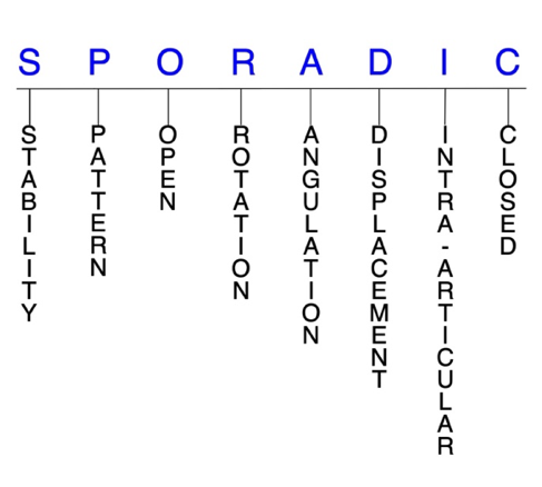
In addition, anatomically named fractures are often also identified by specific eponyms or other special features.

For the Capitate Fracture, the historical and specifically named fractures include:

Scaphocapitate fracture syndrome

Capitate dislocation and fracture-dislocation

By selecting the name (diagnosis), you will be linked to the introduction section of this Diagnostic Guide dedicated to the selected fracture eponym.


Fractures of the carpal bones account for ~6% of fractures overall and up to 18% of all hand fractures. The majority (58-89%) occurs in the scaphoid, while fractures of the other 7 carpals are uncommon and comprise ~1.1% of all fractures. The capitate is the largest and most protected of the carpals; consequently, capitate fractures account for 1-2% of carpal fractures; however, their true incidence is unknown because accurate diagnoses are often missed. Although rare, capitate fractures can be serious injuries that require careful detection and an appropriate management strategy to reduce the risk for complications. Nondisplaced capitate fractures—especially those that are isolated—can often be effectively treated with closed reduction and immobilization, while surgery is typically needed for displaced and/or comminuted fractures, or when conservative methods fail.1-7

Definitions

  • A capitate fracture is a disruption of the mechanical integrity of the capitate.
  • A capitate fracture produces a discontinuity in the capitate contours that can be complete or incomplete.
  • A capitate fracture is caused by a direct force that exceeds the breaking point of the bone.

Hand Surgery Resource’s Fracture Description and Characterization Acronym

SPORADIC

S – Stability; P – Pattern; O – Open; R – Rotation; A – Angulation; D – Displacement; I – Intra-articular; C – Closed


S - Stability (stable or unstable)

  • Universally accepted definitions of clinical fracture stability is not well defined in the hand surgery literature.8-10
  • Stable: fracture fragment pattern is generally nondisplaced or minimally displaced. It does not require reduction, and the fracture fragment’s alignment is maintained with simple splinting. However, most definitions define a stable fracture as one that will maintain anatomical alignment after a simple closed reduction and splinting. Some authors add that stable fractures remain aligned, even when adjacent joints are put to a partial range of motion (ROM).
  • Unstable: will not remain anatomically or nearly anatomically aligned after a successful closed reduction and simple splinting. Typically unstable capitate fractures have significant deformity with comminution, displacement, angulation, and/or shortening.
  • The intercarpal ligaments offer a great deal of extrinsic stability to the capitate, while its cuboidal shape provides additional strength to the bone. This is why the majority of capitate fractures are stable and nondisplaced.3,11

P - Pattern

  • Capitate head: this fracture pattern appears to be rare
  • Capitate neck (waist): the most common site of fracture in the capitate, these injuries are usually transverse in configuration5
  • Capitate body: may be stellate comminuted, oblique, or transverse, but transverse is most common; these fractures can result from a direct blow that causes multiple carpal fractures or as part of an incomplete or self-reduced perilunate injury1,12
  • Other possible capitate fracture patterns include verticofrontal, parasagittal, avulsion tip, or shear depression fractures.2,12
  • The mechanism of injury often dictates the fracture pattern.2
    • Different mechanisms of injury for capitate fractures have been proposed, and the matter is still debated. In many cases, the exact mechanism is difficult to determine.
    • The most common mechanism identified has been a FOOSH with an extended wrist (77%), which applies a dorsiflexion force to the wrist in neutral, ulnar, or radial deviation.
    • A fall onto the dorsum of the hand, which applies a flexion force to the wrist, appears to be the second mechanism in terms of frequency (15.4%).
    • A direct blow or an axial trauma, transmitted through the heads of the index and long metacarpals in a clenched fist and flexed wrist, are also possible causes. Stress fractures and pathological fractures have been described as well.6,7,13
  • Ligaments anchor the body of the capitate to the trapezoid, hamate, and base of the metacarpals, which leaves the head and the neck vulnerable to rapid high-energy impact, bending, axial, shear, and torsion forces. Adjacent carpals or the borders of the radius may impinge the head or neck, further accentuating these forces.5

O - Open

  • Open: a wound connects the external environment to the fracture site. The wound provides a pathway for bacteria to reach and infect the fracture site. As a result, there is always a risk for chronic osteomyelitis. Therefore, open fractures of the capitate require antibiotics with surgical irrigation and wound debridement.8,14,15
  • Open fractures of the capitate may require surgical exploration to determine if articular surfaces are involved. After irrigation and debridement, these wounds are generally left open and further treatment is typically delayed until the wound shows no sign of infection.16,17

R - Rotation

  • Capitate fracture deformity can be caused by rotation of the distal fragment on the proximal fragment.
  • Degree of malrotation of the fracture fragments can be used to describe the fracture deformity.
  • Scaphocapitate fracture syndrome consists of concomitant fractures of the scaphoid and capitate with a rotation of 90-180° of the proximal fragment of the capitate.18
  • Rotatory dislocation of the capitate can be considered a combination of a dorsal carpometacarpal (CMC) joint dislocation of the long metacarpal and a midcarpal dislocation at the luno-capitate articulation.19

A - Angulation (fracture fragments in relationship to one another)

  • Angulation is measured in degrees after identifying the direction of the apex of the angulation.
    • Straight: no angulatory deformity
    • Angulated: bent at the fracture site
      • Example: in displaced oblique capitate neck fractures, the proximal fragment may be ulnarly angulated and pronated in relation to the distal fragment5
  • Due to its anatomic position and inherent stability, isolated capitate fractures are often nondisplaced; however, in perilunate injuries, proximal capitate fragments can be displaced and rotated as much as 180°.5,11,20
  • Since the principal intraosseous blood supply of the capitate flows retrograde from the body to the head, displaced fractures jeopardize the viability of the proximal pole.5

D - Displacement (Contour)

  • Displaced: disrupted cortical contours
  • Nondisplaced: fracture line defining one or several fracture fragments; however, the external cortical contours are not significantly disrupted

I - Intra-articular involvement

  • Fractures that enter a joint with one or more of their fracture lines.
  • Capitate fractures can have fragment involvement with any of its CMC or intercarpal joint articulations.
  • If a fracture line enters a joint but does not displace the articular surface of the joint, then it is unlikely that this fracture will predispose to posttraumatic osteoarthritis. If the articular surface is separated or there is a step-off in the articular surface, then the congruity of the joint will be compromised and the risk of posttraumatic osteoarthritis increases significantly.
  • The implications of displaced intra-articular capitate fractures are the same as fractures of other carpal bones in terms of carpal mechanics.13
  • Dorsal distal articular margin capitate fractures occur as part of long CMC joint fracture-dislocations, where the mechanism of injury is axial loading combined with a flexion moment applied to the long metacarpal.1

C - Closed

  • Closed: no associated wounds; the external environment has no connection to the fracture site or any of the fracture fragments.8-10

Capitate fractures: named fractures, fractures with eponyms and other special fractures

Scaphocapitate fracture syndrome

  • Rare but complex injury that is a manifestation of the perilunate injury pattern.1,21
    • These injuries most commonly occur in young men between ages 20-30.22
    • The most recognized mechanism of injury is a volar-applied force to a hyperextended wrist, such as from a fall from a height or vehicular accident.22
    • This wrist hyperextension results in a scaphoid fracture and, with further extension, the capitate impacts on the dorsal lip of the radius. This produces a transverse capitate body fracture, in which its proximal fragment rotates 90-180° in the sagittal plane as the hand returns to a neutral position. The articular cartilage of the capitate head then faces distally in opposition to the fracture surface of the distal capitate fragment.1
    • Can be either isolated or associated with a perilunate dislocation, but a substantial force is usually required to cause a dislocation.22
  • In one study, scaphocapitate fractures were classified into 6 patterns based on fragment geometry and displacement:
    • Type I: transverse fracture of the scaphoid and capitate without dislocation
    • Type II: inverted proximal fragment of capitate that remains in articulation with the lunate
    • Type III: dorsal perilunate dislocation
    • Type IV: volar perilunate dislocation of the carpus and proximal fragment of capitate
    • Type V: isolated volar dislocation of the proximal fragment of capitate
    • Type VI: isolated dorsal dislocation of the proximal fragment of capitate21
  • Some believe that scaphocapitate fracture syndrome is a variety of trans-scaphoid, trans-capitate, perilunate fracture-dislocation and represents the final stage of a greater arc injury with the potential of spontaneous reduction; however, the different degrees and directions of displacement of the capitate head imply that this injury does not have a unique mechanism.12,18,21

Imaging

  • Plain radiographs may not show the extent of the median nerve lesion. This, combined with the rarity and complexity of these injuries, causes many diagnoses to be initially missed or incorrectly labeled as simple scaphoid fractures.18,21
  • If plain radiography does not lead to a satisfactory diagnosis, a CT scan may be needed, especially if a complex lesion of the carpus is suspected.18,21

Treatment

  • Some controversy exists about the optimal management strategy for a capitate fracture in scaphocapitate fracture syndrome, but conservative treatment—consisting of closed reduction and cast immobilization—may be appropriate for some nondisplaced fractures.21
  • For displaced or comminuted fractures, open reduction and internal fixation (ORIF) is generally considered the treatment-of-choice to reduce complications. The aim of treatment is reduction and fixation of both fractures to obtain bony union.
    • A dorsal approach is most commonly used, while a volar approach is usually reserved for when decompression of the median nerve is necessary.22
    • K-wires or compression screws are typically recommended to achieve fixation and reduce the risk for nonunion, and no significant differences have been identified between these two approaches.18,22
    • According to the literature, it is recommended that reduction of the capitate precedes reduction of the scaphoid.
  • Reduction of the scaphoid fragments is guided from the radial surface of the capitate and is not easily maintained if the capitate is not stabilized, as the proximal scaphoid fragment tends to displace into the gap of the capitate head.18
  • Some authors recommended excision of the displaced proximal fragment of the capitate because of the possibility of avascular necrosis, but it’s been found that the capitate head can revascularize when replaced anatomically and immobilized until fracture healing.21
    • Excision can also interfere with the function of the carpus and eventually result in degenerative arthritis. It is therefore advised that the capitate fragment is not excised, even if it cannot be fixed.21
  • Cancellous bone grafting may be considered in some cases of bone loss.18

Complications

  • Infection
  • Avascular necrosis
  • Nonunion
  • Osteonecrosis

Outcomes

  • For patients with scaphocapitate fracture syndrome, early surgical intervention with meticulous reduction and fixation of all fractures and dislocations present generally leads to a favorable result with minimal complications. Despite the severity of this injury, restoring normal anatomical relationships of the carpus can lead to a successful long-term functional outcome.18
  • In one study of individuals with nondisplaced fractures, those treated surgically returned to work earlier and had better functional outcomes than those treated conservatively.21
  • Delayed diagnosis and treatment of this injury has been found to result in nonunion with subsequent carpal arthritis and carpal collapse.21

Capitate dislocation and fracture-dislocation

  • Dislocation of the capitate in any pattern is extremely rare. When these injuries do occur, it is usually concomitant with other fracture(s) and/or dislocation(s), meaning isolated capitate dislocations and fracture-dislocations are even less common.11,23
    • Transscaphoid perilunate fracture-dislocation and perilunate dislocation are the two major patterns of carpal dislocation, in which the common feature is a dislocation of the head of the capitate from the distal surface of the lunate.19
    • An isolated capitate dislocation is possible when a localized force is concentrated over the bone.23
    • Significant energy is required to fracture and furthermore to displace the fragments in a capitate fracture-dislocation.3
  • Rotatory dislocation of the capitate can be considered a combination of a dorsal CMC dislocation of the long metacarpal and a midcarpal dislocation at the luno-capitate articulation.19

Imaging

  • Radiographic imaging should include posteroanterior (PA), oblique, lateral, and PA with ulnar deviation views.
  • A CT scan may be necessary in some cases to confirm the diagnosis and characterize the orientation of the fracture fragments.

Treatment

  • ORIF followed by immobilization is often necessary for dorsal capitate dislocations. After the immobilization device is removed, rehabilitation to regain wrist ROM should be initiated.23
  • Capitate fracture-dislocations with unsalvageable devascularized fragments and significant ligamentous injury to the carpus may require wrist arthrodesis in which the remaining carpus is fused.3

Complications

  • Infection
  • Avascular necrosis
  • Nonunion

Related Anatomy

  • The capitate consists of a distal body that articulates with the index, long, and ring metacarpals at their respective CMC joints, the trapezoid on the distolateral/radial surface, and the hamate on the medial/ulnar surface, as well as a proximal neck and smooth, rounded head that articulates with the scaphoid and lunate bones at the proximolateral concavity. The dorsal surface is broad and wider than the narrow volar surface, which has a distinct prominence.5,13,20
    • The cuboidal shape of the capitate provides it with inherent strength and makes it the rigid keystone of the carpus that bridges the intersection of the longitudinal and transverse carpal arches. As such, it is integral in the axial movement of the long metacarpal.3,13,20
  • Ligamentous attachments of the capitate include the capitohamate ligament, radiocapitate ligament, radioscaphocapitate ligament, triquetrocapitate ligament, trapeziocapitate ligament, ulnocapitate ligament, a dorsal and volar CMC ligament, and two interosseous ligaments that attach on the lateral and medial surfaces of the capitate body. The head and neck have no ligamentous attachments, which leaves them vulnerable to rapid high-energy impact, bending, axial, shear, and torsion forces.5,20
  • The only tendon associated with the capitate is the oblique head of the adductor pollicis tendon, which arises from several slips of the capitate.5

Incidence and Related injuries/conditions

  • Fractures of the carpal bones have been found to account for between 8-18% of all hand fractures24,25 and ~6% of fractures overall.26
  • Fractures of the proximal carpals are more common than the distal carpals, and the most commonly fractured carpal bone is the scaphoid, which represents 58-89% of all carpal fractures.24,25,27,28
  • Fractures of the other 7 carpals are very rare and only account for ~1.1% of all fractures. The triquetrum is the most commonly involved of these bones, while fractures of the other carpals are even more rare and vary in incidence.29-31
  • Due to its size and stability, capitate fractures have been found to only comprise between 1-2% of all carpal fractures;11,32,33 however, the true incidence of these injuries is not clearly known, as the diagnosis is often missed.7
    • About 50% of capitate fractures are associated with concomitant osseous and/or ligamentous injuries, while the other 50% are isolated fractures.1
  • One study found the incidence of isolated capitate fractures to be 0.3% of all carpal fractures, as most fractures are associated with additional wrist pathology like perilunate injuries and scaphoid fractures.7,32
  • Another study found that 57% of initial X-rays failed to reveal an isolated capitate fracture, which shows why the reported incidence of these injuries is likely underestimated.6
    • Capitate fractures occur most frequently in younger male patients between ages 20-30 who may be more prone to high-energy trauma than the general population. For this reason, coincident polytrauma is frequent in this population.5,7

Work-up Options

  • Routine X-rays
    • Standard wrist radiographs may not reveal isolated body or dorsal articular margin capitate fractures, especially if the fracture is nondisplaced. This is one of the main reasons many capitate fractures are undiagnosed.1,5,20
  • Special X-ray views
    • The dorsal tilt view, which is a PA projection with the central beam angled 25-30° towards the fingers and centered on the capitate, is useful when there is a suspicion of capitate neck fracture.34
    • Radial and ulnar deviation views in the anteroposterior (AP) plane are also helpful for making capitate fractures more evident.7
    • Carpal tunnel view34
  • CT scan
    • May be necessary for occult capitate fractures that don’t appear on plain radiographs. In these cases, a CT scan in the coronal plane for transverse fractures and in the parasagittal plane for articular margin fractures is recommended.1,5,11
  • MRI
    • May also be needed for occult capitate fractures.5,11,12
    • MRIs are helpful for achieving a prompt diagnosis and appropriate treatment regimen, and for delineating fracture displacement and associated ligament injury.2,7
    • MRIs are also useful for predicting healing potential of transverse body fractures by visualizing the vascular status of the capitate head.1
  • Isotope bone scan
    • Another imaging option for occult capitate fractures when radiographs are inconclusive.5,11
ICD-10 Codes
  • CAPITATE FRACTURE

    Diagnostic Guide Name

    CAPITATE FRACTURE

    ICD 10 Diagnosis, Single Code, Left Code, Right Code and Bilateral Code

    DIAGNOSISSINGLE CODE ONLYLEFTRIGHTBILATERAL (If Available)
    FRACTURE CARPAL BONE - CAPITATE    
    - DISPLACED S62.132_S62.131_ 
    - NONDISPLACED S62.135_S62.134_ 

    Instructions (ICD 10 CM 2020, U.S. Version)

    THE APPROPRIATE SEVENTH CHARACTER IS TO BE ADDED TO EACH CODE FROM CATEGORY S62
     Closed FracturesOpen Type I or II or OtherOpen Type IIIA, IIIB, or IIIC
    Initial EncounterABC
    Subsequent Routine HealingDEF
    Subsequent Delayed HealingGHJ
    Subsequent NonunionKMN
    Subsequent MalunionPQR
    SequelaSSS

    ICD-10 Reference

    Reproduced from the International statistical classification of diseases and related health problems, 10th revision, Fifth edition, 2016. Geneva, World Health Organization, 2016 https://apps.who.int/iris/handle/10665/246208

Clinical Presentation Photos and Related Diagrams
Capitate Fracture Diagrams
  • Transverse fracture (arrow) of the body of the capitate AP view
    Transverse fracture (arrow) of the body of the capitate AP view
  • Transverse fracture (curved arrow) of the body of the capitate lateral view with double arrow on capitate outline
    Transverse fracture (curved arrow) of the body of the capitate lateral view with double arrow on capitate outline
  • Fracture (arrow) of the dorsal lip of the capitate AP view
    Fracture (arrow) of the dorsal lip of the capitate AP view
  • Fracture (curved arrow) of the dorsal lip of the capitate lateral view with double arrow on capitate outline
    Fracture (curved arrow) of the dorsal lip of the capitate lateral view with double arrow on capitate outline
  • Transverse fracture (arrow) of the neck of the capitate AP view
    Transverse fracture (arrow) of the neck of the capitate AP view
  • Fracture (curved arrow) of the neck of the capitate lateral view with double arrow on capitate outline.  Capitate head at risk for AVN with this fracture
    Fracture (curved arrow) of the neck of the capitate lateral view with double arrow on capitate outline. Capitate head at risk for AVN with this fracture
Symptoms
History of trauma
Fracture pain and deformity
Wrist swelling and ecchymosis
Typical History

A typical patient is a 23-year-old, left-handed man who injured his right wrist after falling from the handlebars of a bike. The man was riding on his friend’s handlebars at a fast speed when a car suddenly emerged from an alleyway. In response, the friend slammed on the brakes, which launched him several feet forward. The man landed directly onto his outstretched hands, with the right hand taking the brunt of the impact. The accident produced a flexion force on his right wrist and fractured the capitate and scaphoid bones, and dislocated 2 of the capitate’s articulations.

Positive Tests, Exams or Signs
Work-up Options
Treatment Options
Treatment Goals
  • When treating closed capitate fractures, the treating surgeon has 4 basic goals:8,15
    1. A wrist with a normal appearance. The X-ray may not need to be perfect, but the capitate should have no obvious deformity (ie, the capitate looks normal!)
    2. Avoid stiffness by maintaining a normal functional ROM (ie, the wrist works!)
    3. The capitate is not painful (ie, the capitate does not hurt!)
    4. Congruent joint surface with none-to-minimal joint surface irregularities (ie, the joints associated with the capitate do not develop early posttraumatic arthritis!)
    5. Fracture care should minimize the risk for infection and osteomyelitis.
  • One additional goal is mandatory for open fractures:
Conservative
  • The management of capitate fractures is challenging and is based on the fracture pattern/location and the presence of other related injuries.2,12
  • The majority of capitate fractures occur in conjunction with other osseous and ligamentous injuries to the carpus, but because of the bone’s size, shape, and location, they are usually stable and non-displaced.10,17
    • Most capitate fractures that are closed, nondisplaced, and minimally angulated without significant malrotation can be managed with closed reduction and immobilization using a cast or aluminum, plaster, fiberglass, or custom splint.10,12,17,35
      • This treatment approach is particularly applicable to the rare isolated capitate fracture.1
      • Immobilization of these injuries is recommended for 6-12 weeks.7
      • Exercises to maintain ROM of the shoulder, thumb, and fingers may begin immediately, while a more intense rehabilitation protocol should be initiated as soon as the immobilization device is removed.7
  • Capitate head fractures are entirely covered with articular cartilage, and their intrasynovial location may delay union. Therefore, conservative treatment may not be appropriate in these cases.2
  • Even capitate fractures that require a reduction to correct fracture-related deformity usually can be held in anatomic or near-anatomic alignment with a splint without internal or external surgical fixation.17
Operative
  • Surgical treatment of capitate fractures must always be an individualized therapeutic decision. However, surgical capitate fracture care is most frequently recommended when:
    1. Closed reduction fails or the simple splint or cast immobilization does not maintain the reduction. For these irreducible or unstable fractures, operative treatment is recommended to achieve the 4 treatment goals of fracture care.
    2. There is a significantly displaced capitate fracture fragment involving one of its associated joints. Surgical fracture care may be required in these cases.
    3. Open capitate fractures require surgical care in the form of irrigation and debridement to prevent chronic infection.
  • For displaced and/or comminuted capitate fractures, ORIF is typically recommended.
    • Internal fixation can be achieved with either K-wires or headless compression screws.7,12
  • Positive outcomes have been identified with both fixation devices, but screws have the advantage over K-wires of providing compression across the fracture site and allowing early motion. For this reason, some authors recommend screws over K-wires.2,11
    • Despite devascularization of the proximal fragment, ORIF of displaced capitate fractures may result in fracture healing and revascularization of the proximal fragment in some patients.5,11
    • Capitate fractures are usually most readily accessible through a dorsal incision, although combined dorsal and volar incisions have also been used.5,11
    • For capitate neck fractures, cast immobilization alone may extend for several months with potential concomitant wrist stiffness, while rigid internal fixation is more likely to expedite treatment.2
  • Capitate fractures that are part of a larger wrist injury should be fixed along with associated osseous/ligamentous injuries.1
  • Fractures that are apparently isolated but displaced require meticulous evaluation, as displacement can occur with concomitant osseous or ligamentous injuries.1
    • Increasing displacement of capitate fractures risk disruption of the tenuous blood supply, since the capitate is perfused in a retrograde fashion, with 67% of the vessels entering dorsally.3
  • Other surgical interventions for capitate fractures and associated complications—like avascular necrosis and nonunion—include surgical excision, midcarpal arthrodesis, bone grafting, partial resection, or arthroplasty with silicone; however, reports on the effectiveness of these techniques are lacking in the literature.7

Post-treatment Management

  • The care and precautions related to immobilization devices for the capitate fracture must be carefully reviewed with the patient. Patients should be educated regarding care and precautions. Patients should know that pain, especially increasing pain, numbness, tingling, skin irritation, splint loosening, or excessive splint tightness are red flags and should be reported to the surgeon or his team.
  • Pain should be managed with properly fitting splints, reassurance, elevation, ice in the initial post-fracture period, and mild pain medications. Patients should be encouraged to discontinue pain medication as soon as possible. Opioid use should be kept to a minimum.
  • Patients should be instructed to carefully exercise all joints in the injured hand and arm that do not require immobilization. Patients usually can exercise on their own; however, signs of generalized finger or hand stiffness are indications for referral to hand therapy (PT or OT).
    • After surgery, a cast or splint is used for immobilization, and once removed, rehabilitation may begin. Treatment is intended to restore hand and wrist function, and exercises that utilize therapeutic putty, hand therapy balls, and grip-strengthening devices can be used to strengthen muscles around the wrist and alleviate swelling.3,23
  • If an infection does occur, management should focus on eradicating sepsis with thorough debridement, appropriate antibiotics (eg, cephalosporin, penicillin), and fracture stabilization, followed by obtaining fracture union and regaining a functional extremity.16
Complications
  • The complication rate for capitate fractures is relatively high and the associated consequences of these complications are important to recognize.6
  • Nonunion has been identified as the most common of all complications, reported in 19.6-56% of isolated capitate fractures.3,11
    • Nonunion most commonly results from delay in diagnosis and lack of initial treatment, and it is associated with absorption of the fracture surfaces and shortening of the capitate, which induces a collapse deformity.3,6
  • The retrograde blood supply pattern is also partially responsible for nonunion.12
    • This complication can be treated with intercalary bone grafting and/or ORIF, by partial resection and/or carpal fusion, or by wrist arthrodesis.
  • Without treatment of nonunion, pain and disability in the wrist may persist that is similar to chronic wrist sprain associated with untreated scaphoid fractures and carpal tunnel syndrome.6
  • Avascular necrosis—especially of the proximal pole—is another common complication that is frequently seen in displaced capitate fractures.
    • This complication occurs because the capitate head has no soft-tissue attachments and because of the retrograde pattern of blood supply in the bone, which renders the proximal capitate avascular in cases of capitate body fracture.12
    • Fractures of the head or neck interrupt the intraosseous circulation to the proximal fragment, and the peripheral blood flow of the proximal ligament is insufficient for reliable fracture healing. The more proximal these fractures, the greater these risks.5
      • Fortunately, the avascular proximal end of the capitate can be revascularized if reduced anatomically and immobilized until healing is complete.6
    • Other possible complications with capitate fractures are stiffness, dorsiflexion instability, capitate collapse, midcarpal arthrosis, capitolunate fusion, posttraumatic arthritis, and malunion.2,6,12
      • Patients with capitate fractures should be counseled that functional limitation is common secondary to nonunion, avascular necrosis of the proximal pole, capitate collapse, symptomatic midcarpal arthrosis, or associated injuries.
      • Painful midcarpal arthrosis can be treated with midcarpal arthrodesis.1
    • Chronic osteomyelitis of the capitate is rare but can occur in open fractures, especially in diabetic patients or those with a compromised immune system.
Outcomes
  • In general, most outcomes for capitate fractures are positive,12,16-18,21 but isolated capitate fracture outcomes have not been as clearly defined.6
  • In one trial, 11 patients with an isolated capitate fracture were initially treated conservatively.
    • Two patients subsequently required surgery due to persistent pain and delayed healing.
    • All patients were followed-up a median of 7.9 months after presentation, and at the latest follow-up, 10 patients had functional wrist ROM.
  • Three patients reported pain, including the 2 patients who had surgery.
  • One of these patients had findings of early midcarpal arthrosis and the other developed a nonunion of the capitate that required revision surgery.2
Key Educational Points
  • The majority of capitate fractures can be treated without surgery.10,16
  • Underlying pathological conditions such as bone tumors—like enchodromas—and osteoporosis should be expected in fractures that occur from trivial trauma.
  • The functional needs of each patient must be considered when recommending treatment for capitate fractures.
  • The most common pattern of associated injury with capitate fractures is a transscaphoid, transcapitate, greater arc perilunate injury. The biomechanics of this injury pattern relate to the scaphoid’s role as the connecting link between the 2 carpal rows, with the lunocapitate joint acting as a junction between the proximal and the distal carpal rows.12
  • With regular MRI examination, the diagnosis of capitate fractures can be confirmed and the evolution can be followed. When bone circulation remains compromised or when the healing seems to slow down, operative treatment should be considered.6
References

New and Cited Articles

  1. Shah MA, Viegas SF. Fractures of the carpal bone excluding the scaphoid. J Hand Surg Am 2002; 2(3): 129-140
  2. Suh N, Ek ET, Wolfe SW. Carpal fractures. J Hand Surg Am 2014;39(4):785-91. PMID: 24679911
  3. Ting M, Kusnezov N, Dunn JC, et al. Pancarpal dissociation with palmar Capitate fracture-dislocation: Case report. Orthop Traumatol Surg Res 2017;103(3):457-459. PMID: 28274880
  4. Murthy NS. The role of magnetic resonance imaging in scaphoid fractures. J Hand Surg Am 2013;38(10):2047-54. PMID: 24079527
  5. Freeland AE, Pesut TA. Oblique capitate fracture of the wrist. Orthopedics 2004;27(3):287-90. PMID: 15058451
  6. De Schrijver F, De Smet L. Isolated fracture of the capitate: the value of MRI in diagnosis and follow up. Acta Orthop Belg 2002;68(3):310-5. PMID: 12152383
  7. Palumbo DM. An uncommon isolated fracture of the capitate bone. JAAPA 2007;20(12):33-5. PMID: 18181367
  8. Cheah AE, Yao J. Hand Fractures: Indications, the Tried and True and New Innovations. J Hand Surg Am 2016;41:712-22. PMID: 27113910
  9. Nesbitt KS, Failla JM, Les C. Assessment of instability factors in adult distal radius fractures. J Hand Surg Am 2004;29:1128-38. PMID: 15576227
  10. Walenkamp MM, Vos LM, Strackee SD, Goslings JC, Schep NW. The Unstable Distal Radius Fracture-How Do We Define It? A Systematic Review. J Wrist Surg 2015;4:307-16. PMID: 26649263
  11. Sabat D, Arora S, Dhal A. Isolated capitate fracture with dorsal dislocation of proximal pole: a case report. Hand (N Y) 2011;6(3):333-6. PMID: 22942861
  12. Kadar A, Morsy M, Sur YJ, et al. Capitate Fractures: A Review of 53 Patients. J Hand Surg Am. 2016;41(10):e359-e366. PMID: 27524693
  13. Richards RR, Paitich CB, Bell RS. Internal fixation of a capitate fracture with Herbert screws. J Hand Surg Am 1990;15(6):885-7. PMID: 2269778
  14. Ketonis C, Dwyer J, Ilyas AM. Timing of Debridement and Infection Rates in Open Fractures of the Hand: A Systematic Review. Hand (N Y) 2017;12:119-26. PMID: 28344521
  15. Meals C, Meals R. Hand fractures: a review of current treatment strategies. J Hand Surg Am 2013;38:1021-31. PMID: 23618458
  16. Day CS. Fractures of the Metacarpals and Phalanges. In: Green DP, ed. Green's Operative Hand Surgery. Seventh ed. Philadelphia: Elsevier; 2016, pp. 231-77.
  17. Weinstein LP, Hanel DP. Metacarpal fractures. J Hand Surg Am 2002; 2(4):168–180.
  18. Dailiana ZH, Papatheodorou LK, Malizos KN. Scaphocapitate Fracture: Two Cases with Follow-Up over 5 Years. J Wrist Surg 2015;4(3):174-8. PMID: 26261742
  19. Hirata H, Sasaki H, Ogawa A, et al. Rotary dislocation of the capitate: a case report. J Hand Surg Am 1997;22(1):89-90. PMID: 9018619
  20. Robbins MM, Nemade AB, Chen TB, Epstein RE. Scapho-capitate Syndrome Variant: 180-Degree Rotation of the Proximal Capitate Fragment Without Identifiable Scaphoid Fracture. Radiol Case Rep 2015;3(3):193. PMID: 27303543
  21. Kim YS, Lee HM, Kim JP. The scaphocapitate fracture syndrome: a case report and literature analysis. Eur J Orthop Surg Traumatol 2013;23 Suppl 2:S207-12. PMID: 23412323
  22. Hamdi MF. The scaphocapitate fracture syndrome: report of a case and a review of the literature. Musculoskelet Surg 2012;96(3):223-6. PMID: 21373909
  23. Rosh AJ, Schwartz DT. Isolated capitate and hamate dislocation. J Emerg Med 2012;42(6):e151-2. PMID: 20800413
  24. van Onselen EB, Karim RB, Hage JJ, Ritt MJ. Prevalence and distribution of hand fractures. J Hand Surg Br 2003;28(5):491-5. PMID: 12954264
  25. Hove LM. Fractures of the hand. Distribution and relative incidence. Scand J Plast Reconstr Surg Hand Surg 1993;27(4):317-9. PMID: 8159947
  26. Dobyns JH, Beckenbaugh RD, Bryan RS, et al. Fractures of the hand and wrist. In: Flynn JE, editor. Hand surgery. Third ed. Philadelphia: Lippincott Williams & Wilkins; 1982.
  27. Rhemrev SJ, Ootes D, Beeres FJ, et al. Current methods of diagnosis and treatment of scaphoid fractures. Int J Emerg Med 2011;4:4. PMID: 21408000
  28. Hey HW, Chong AK, Murphy D. Prevalence of carpal fracture in Singapore. J Hand Surg Am 2011;36(2):278-83. PMID: 21276891
  29. Larsen CF, Brøndum V, Skov O. Epidemiology of scaphoid fractures in Odense, Denmark. Acta Orthop Scand 1992;63(2):216-8. PMID: 1590062
  30. Höcker K, Menschik A. Chip fractures of the triquetrum. Mechanism, classification and results. J Hand Surg Br 1994;19(5):584-8. PMID: 7822914
  31. Garcia-Elias M.Dorsal fractures of the triquetrum-avulsion or compression fractures? J Hand Surg Am 1987;12(2):266-8. PMID: 3559084
  32. Rand JA, Linscheid RL, Dobyns JH. Capitate fractures: a long-term follow-up. Clin Orthop Relat Res 1982;(165):209-16. PMID: 7075062
  33. Apostolides JG, Lifchez SD, Christy MR. Complex and rare fracture patterns in perilunate dislocations. Hand (N Y) 2011;6(3):287-94. PMID: 22942853
  34. Bhat AK, Kumar B, Acharya A. Radiographic imaging of the wrist. Indian J Plast Surg 2011;44(2):186-96. PMID: 22022028
  35. Kollitz KM, Hammert WC, Vedder NB, Huang JI. Metacarpal fractures: treatment and complications. Hand (N Y) 2014;9(1):16-23. PMID: 24570632

Reviews

  1. Papp S.Carpal bone fractures. Hand Clin 2010;26(1):119-27. PMID: 20006250
  2. Pan T, Lögters TT, Windolf J, Kaufmann R. Uncommon carpal fractures. Eur J Trauma Emerg Surg 2016;42(1):15-27. PMID: 26676306

Classics

  1. Brekkan A, Karlsson J, Thorsteinsson T. Case report 252. Scapho-capitate fracture of the right wrist with dislocation and rotation to 180 degrees of the proximal fragment of the capitate. Skeletal Radiol 1983;10(4):291-3. PMID: 6648570
  2. Monahan PR, Galasko CS. The scapho-capitate fracture syndrome. A mechanism of injury. J Bone Joint Surg Br 1972;54(1):122-4. PMID: 5011738
Subscribe to CAPITATE FRACTURE