Introduction

Fracture Nomenclature for Pediatric Capitellar Fractures

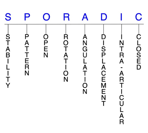
Hand Surgery Resource’s Diagnostic Guides describe fractures by the anatomical name of the fractured bone and then characterize the fracture by the Acronym:
In addition, anatomically named fractures are often also identified by specific eponyms or other special features.

For the Pediatric Capitellar Fractures, the historical and specifically named fractures include:

Kocher-Lorenz Fracture

By selecting the name (diagnosis), you will be linked to the introduction section of this Diagnostic Guide dedicated to the selected fracture eponym.


Fractures of the capitellum are rare in adults, and even less common in children and adolescents. The most common mechanism of injury for pediatric capitellar fractures is a fall on an outstretched hand with the elbow extended or semi-flexed, which results in a shearing force across the capitellum that displaces a fragment proximally. Since the capitellum is primarily cartilaginous prior to age 12, the lateral condyle is more likely to fracture from this mechanism than the capitellum. Due to their low incidence, capitellar fractures are often misdiagnosed or not treated appropriately, which can adversely affect long-term outcomes. Conservative treatment using cast or splint immobilization is indicated for stable and nondisplaced fractures, while surgery may be indicated for more severe fracture patterns and those that cannot be reduced nonsurgically.1-4

Definitions

  • A pediatric capitellar fracture is a disruption of the mechanical integrity of the capitellum.
  • A pediatric capitellar fracture produces a discontinuity in the capitellum contours that can be complete or incomplete.
  • A pediatric capitellar fracture is caused by a direct force that exceeds the breaking point of the bone.

Hand Surgery Resource’s Fracture Description and Characterization Acronym

SPORADIC

S – Stability; P – Pattern; O – Open; R – Rotation; A – Angulation; D – Displacement; I – Intra-articular; C – Closed


S - Stability (stable or unstable)

  • Universally accepted definitions of clinical fracture stability are not well defined in the literature.5-7
  • Stable: fracture fragment pattern is generally nondisplaced or minimally displaced. It does not require reduction, and the fracture fragments’ alignment is maintained by with simple splinting or casting. However, most definitions define a stable fracture as one that will maintain anatomical alignment after a simple closed reduction and splinting. Some authors add that stable fractures remain aligned, even when adjacent joints are put to a partial range of motion (ROM).
  • Unstable: will not remain anatomically or nearly anatomically aligned after a successful closed reduction and immobilization. Typically unstable pediatric capitellar fractures have significant deformity with comminution, displacement, angulation, and/or shortening.

P - Pattern2,8,9

  • Type I (Hahn-Sternthal): shear fracture involving most of the capitellum and little or none of the trochlea; the fracture fragment has a significant bony component; this pattern is common in children
  • Type II (Kocher-Lorenz): osteochondral fracture involving a variable amount of articular cartilage of the capitellum with minimal attached subchondral bone; this pattern is very rare in children
  • Type III (Broberg-Morrey): comminuted or compression fracture of the capitellum
  • Type IV (McKee): shear coronal fracture of the distal humerus involving the capitellum and a large portion of the trochlea

O - Open

  • Open: a wound connects the external environment to the fracture site. The wound provides a pathway for bacteria to reach and infect the fracture site. As a result, there is always a risk for chronic osteomyelitis. Therefore, open fractures of the capitellum require antibiotics with surgical irrigation and wound debridement.5,10,11

R - Rotation

  • Pediatric capitellar fracture deformity can be caused by proximal rotation of the fracture fragment in relation to the distal fracture fragment.
  • Degree of malrotation of the fracture fragments can be used to describe the fracture deformity.
  • Fracture fragments in pediatric capitellar fractures are typically rotated internally.12

A - Angulation (fracture fragments in relationship to one another)

  • Angulation is measured in degrees after identifying the direction of the apex of the angulation.
  • Straight: no angulatory deformity
  • Angulated: bent at the fracture site

D - Displacement (Contour)

  • Displaced: disrupted cortical contours
  • Nondisplaced: ≥1 fracture lines defining one or several fracture fragments; however, the external cortical contours are not significantly disrupted
  • Fracture fragments in pediatric capitellar fractures are typically displaced proximally.12

I - Intra-articular involvement

  • Intra-articular fractures are those that enter a joint with ≥1 of their fracture lines.
  • All pediatric capitellar fractures are considered intra-articular fractures.12
  • Isolated capitellar fractures can have fragment involvement with the radiocapitellar joint, while concomitant fractures with the trochlea can also involve the ulnohumeral joint.
  • If a fracture line enters a joint but does not displace the articular surface of the joint, then it is unlikely that this fracture will predispose to post-traumatic osteoarthritis. If the articular surface is separated or there is a step-off in the articular surface, then the congruity of the joint will be compromised, and the risk of post-traumatic osteoarthritis increases significantly.

C - Closed

  • Closed: no associated wounds; the external environment has no connection to the fracture site or any of the fracture fragments.4-6

Pediatric capitellar fractures: named fractures, fractures with eponyms and other special fractures


Kocher-Lorenz Fracture

  • In the pediatric population, type II capitellar fractures are referred to as the Kocher-Lorenz fracture.9
  • These are chondral shear fractures in which the fractured fragment consists almost entirely of cartilage, with minimal or no subchondral bone. The mechanism of injury is a shear force upon the capitellum by the radius, usually from a fall on a partially flexed elbow, which shears off the chondral fragment from the anterior capitellum.9
  • Kocher-Lorenz fractures are extremely rare, particularly in younger children, because the capitellum is still primarily cartilaginous and thus very resistant to shearing stresses.9
  • Fractured fragments are not visible on plain radiographs, and these injuries are therefore often missed in the acute phase.9

Imaging

  • Radiology studies - X-ray
    • X-rays are often performed initially, and the osteochondral shell of the capitellum should be thoroughly investigated.13
  • Magnetic resonance imaging - MRI without contrast
    • Since plain radiography is typically normal, MRI is needed to accurately diagnose these injuries.9

Treatment

  • Surgery is typically required for Kocher-Lorenz fractures and operative management depends on the duration of injury.9
    • Repositioning and fixation of the fracture fragment is possible if the injury is diagnosed early, and fixation options include K-wires, headless screws, and bio-absorbable screws.
    • When the diagnosis is delayed, the fracture fragment may become hypertrophied and misshapen, which often necessitates fragment excision.

Complications

  • Loss of elbow ROM
  • Stiffness
  • Infection
  • Osteoarthritis

Outcomes

  • Since Kocher-Lorenz fractures are extremely rare, outcome data is scarce and limited to case reports.9

Related Anatomy4,12,14-16

  • The elbow is a hinge-type synovial joint comprised of the radius, ulna, and humerus, and formed by three articulations: the ulnohumeral joint, radiocapitellar joint, and proximal radioulnar joint.
  • The ulnohumeral joint is a hinge joint in which the trochlear notch (or semilunar notch) of the ulna articulates with the trochlea of the humerus. This joint allows for elbow flexion and extension.
    • The trochlea is the medial portion of the articular surface of the distal humerus, which is contained between the lateral and medial columns of the elbow and is primarily covered with articular cartilage. It has medial and lateral ridges with an intervening trochlear groove.
  • The radiocapitellar joint is the articulation of the radial head with the capitellum of the humerus. It is essential to elbow longitudinal and valgus stability and has an integral relationship with the lateral collateral ligament (LCL).
    • The capitellum is a smooth, round, hemispheric structure that represents a portion of a forward-and downward-projecting sphere and which forms the anterior and inferior articular surface of the distal humerus. It is covered with articular cartilage on its anterior and inferior sides, but not its posterior side.
    • The elbow’s axis of flexion-extension and the axis of forearm rotation both pass through the capitellum, which enables effective reach by allowing the hand to function at different distances from the body.
  • The capitellum ossifies around 1 year of age and the lateral portion of the trochlea ossifies at 7 years. The capitellum and trochlea usually fuse at 12 years, but this may occur as early as 9 years. This combined ossification center fuses with the lateral epicondyle around this time to form the main body of the distal humeral epiphysis, which attaches to the metaphysis of the humerus between 12–13 years of age.
  • The key ligaments of the elbow include the LCL (which extends from the lateral epicondyle and blends with the annular ligament of the radius), the medial collateral ligament (MCL, which originates from the medial epicondyle and attaches to the coronoid process and olecranon of the ulna), and the annulus ligament of the radius (which encircles the radial head and stabilizes the radial notch).
  • The key tendons of the elbow include the tendons associated with the biceps, triceps, extensor carpi radialis brevis (ECRB), and extensor carpi radialis longus (ECRL) muscles.

Incidence

  • Capitellar fractures are extremely rare in the pediatric population. They have been found to account for <1% of all pediatric elbow fractures, with one study identifying only 1 capitellar fracture in a series of 2,000 elbow fractures in children.4,9
  • Pediatric capitellar fractures occur predominantly in adolescents, with one study reporting an average age of 14.7 years and a range of 11–17 years. These fractures also appear to be more common in female than male patients.4,9
    • Pediatric capitellar fractures rarely occur in children younger than 12 years, as the mechanism of injury will typically produce a supracondylar fracture due to the highly cartilaginous composition of the capitellum at this age.13

ICD-10 Codes
  • CAPITELLAR FRACTURE - PEDIATRIC

    Diagnostic Guide Name

    CAPITELLAR FRACTURE - PEDIATRIC

    ICD 10 Diagnosis, Single Code, Left Code, Right Code and Bilateral Code

    DIAGNOSISSINGLE CODE ONLYLEFTRIGHTBILATERAL (If Available)

    Instructions (ICD 10 CM 2020, U.S. Version)

    THE APPROPRIATE SEVENTH CHARACTER IS TO BE ADDED TO EACH CODE FROM CATEGORY
     Closed FracturesOpen Type I or II or OtherOpen Type IIIA, IIIB, or IIIC
    Initial EncounterABC
    Subsequent Routine HealingDEF
    Subsequent Delayed HealingGHJ
    Subsequent NonunionKMN
    Subsequent MalunionPQR
    SequelaSSS

    ICD-10 Reference

    Reproduced from the International statistical classification of diseases and related health problems, 10th revision, Fifth edition, 2016. Geneva, World Health Organization, 2016 https://apps.who.int/iris/handle/10665/246208

Symptoms
History of trauma
Fracture pain
Fracture deformity
Swelling, ecchymosis & tenderness
Abrasion
Typical History

A typical patient is a 14-year-old boy who was injured while riding a motorized dirt bike. The boy attempted to ride his bike off a small jump but landed too far forward on the front wheel and tumbled over the handlebars. He landed with both hands outstretched and his elbows extended, with the left arm bearing the brunt of the impact. The landing produced a shearing force on the capitellum and fractured the bone, which led to pain and inflammation in the elbow. The boy was taken to the emergency room soon thereafter.

Positive Tests, Exams or Signs
Work-up Options
Treatment Options
Treatment Goals
  • When treating closed pediatric capitellar fractures, the treating surgeon has 4 basic goals:5,11
    1. An elbow with a normal appearance. The X-ray may not need to be perfect, but the elbow should have no obvious deformity (ie, the elbow looks normal!)
    2. Avoid elbow stiffness by maintaining a normal functional ROM (ie, the elbow works!)
    3. The elbow is not painful (ie, the elbow does not hurt!)
    4. Congruent joint surface with none-to-minimal joint surface irregularities (ie, the elbow does not develop early post-traumatic arthritis!)
  • One additional goal is mandatory for open fractures:
    1. Fracture care should minimize the risk for infection and osteomyelitis.
Conservative
  • Treatment decisions for pediatric capitellar fractures should be individualized according to fracture type, degree of comminution, presence of associated elbow injuries, and the time elapsed since the injury.2
  • Most nondisplaced, stable pediatric capitellar fractures can be treated non-surgically with a period of immobilization in a cast or splint.2,4
Operative
  • Surgical treatment of pediatric capitellar fractures must always be an individualized therapeutic decision. 4,13,19  However, operative capitellar fracture care is most frequently recommended when:
    1. Closed reduction fails or the simple splint or cast immobilization does not maintain the reduction. For these irreducible or unstable fractures, operative treatment is recommended to achieve the 4 treatment goals of fracture care.
    2. There is a significantly displaced trochlear fracture involving the radiocapitellar or ulnohumeral joint.
    3. Open capitellar fractures. These injuries require surgical care in the form of irrigation and debridement to prevent chronic infection.
  • Open reduction and internal fixation (ORIF)
    • Typically indicated for most type I and type IV fractures that warrant surgical intervention.
    • Fixation methods include smooth K-wires, bioabsorbable or cannulated headless compression screws, lag screws, and bioabsorbable pins.
      • K-wires have historically been the preferred fixation method, but wires may not provide stable fixation, and cast immobilization for an extended period is required.
      • Cannulated screws provide sufficient interfragmentary reduction and compression.
    • A lateral extensile approach is preferred, as it helps to view the most medial extension of the fracture.
  • Fragment excision
    • Typically recommended for most type II and type III fractures that require surgery and for small fracture fragments.
  • Closed reduction and percutaneous pinning (CRPP)
    • Less frequently utilized for these injuries. May include the use of a Stienmann pin.
    • Large cartilaginous fragments might prevent proper visualization during this procedure.

Post-treatment Management

  • The care and precautions related to immobilization devices for the capitellar fracture must be carefully reviewed with the patient. Patients should be educated regarding care and precautions. Patients should know that pain, especially increasing pain, numbness, tingling, skin irritation, splint loosening, or excessive tightness are red flags and should be reported to the surgeon or his team.
  • Pain should be managed with properly fitting splints and casts, reassurance, elevation, ice in the initial post-fracture period, and mild pain medications. Patients should be encouraged to discontinue pain medication as soon as possible. Opioid use should be kept to a minimum.
  • Joints that are splinted for closed stable fractures are usually immobilized.
  • Fractures that require internal fixation can be mobilized after 4 weeks.
  • After surgery, the elbow is splinted in a stable position for 2 weeks, after which the patient can begin moving the joint. The integrity of the LCL and MCL should be evaluated and used to create a ligament-specific rehabilitation protocol. Unrestricted ROM can begin at 6 weeks, and then progressive strengthening at 8–10 weeks after surgery so long as there is evidence of fracture healing.17
Complications

Complications13,17,18,20

  • Osteonecrosis
  • Malunion
  • Nonunion
  • Stiffness
  • Post-traumatic osteoarthritis
  • Avascular necrosis
  • Fixation failure
  • Osteomyelitis
  • Instability
  • Infection
Outcomes
  • Since pediatric capitellar fractures are rare, treatment outcome data is scarce.3,18
  • Fragment excision is considered a simple procedure for most displaced type II fractures that has been shown to permit an early return of function in children.2
  • ORIF followed by early mobilization has also been associated with excellent outcomes.4
  • In a series of seven adolescent patients with capitellar fractures, five were treated surgically and all experienced good results. All patients achieved full pronation and supination, while only three had full flexion and extension.3
  • In another series of 9 published case reports and case series of pediatric patients with capitellar fractures, ORIF for displaced anterior shear fractures was generally associated with good results.18
  • Case reports and case series have also shown that patients with Type I and Type II nondisplaced fractures treated nonsurgically with immobilization experienced excellent outcomes.18
Key Educational Points
  • Healthcare providers must investigate for concomitant injury during the diagnosis of pediatric capitellar fractures, such as trochlear fractures, LCL or MCL lesions, or ipsilateral fractures (eg, radial head fractures or epicondylar humeral fractures), which can have significant implications on treatment decisions.21
  • Pediatric capitellar fractures can lead to a reduction in the arm’s arc of motion. If this produces a loss of flexion of >30°, it will adversely impact the reach of the arm and can lead to a contracture from scarring of the soft tissues.15
  • In young children with open physes, surgeons must ensure that the posterolateral soft tissue sleeve of the elbow is left intact during the surgery, as damaging it can interfere with the vascular supply to the lateral column of the distal humerus.4
  • Pediatric capitellar fractures are frequently misdiagnosed, but clinicians must acknowledge that these are rare, distinct injuries that require a different treatment approach than the more frequently seen lateral condyle fractures. Accurate diagnosis is therefore imperative.9,13
  • The ideal surgical approach for pediatric capitellar fractures is yet to be clearly defined. ORIF is currently favored, although fragment excision was once the preferred surgical technique.2,3
  • Anteroposterior (AP), lateral, oblique, and radiocapitellar views of the elbow are necessary.17
  • Pediatric capitellar fractures are often difficult to identify and can be easily missed on radiographs. Contralateral radiographs are therefore recommended, as are true lateral views, which are extremely helpful. Oblique views are typically only helpful if an osseous fragment is present.4,16
  • The “double arc” sign—which represents the displaced capitellum and lateral trochlea—is often visible on a lateral radiographic view and is characteristic of type IV capitellar fractures.4
  • CT scanning can help to classify the fracture by delineating its full extent.4
  • Magnetic resonance imaging - arthrogram may be particularly helpful in this population.4
  • Magnetic resonance imaging - MRI without contrast may be helpful for confirming the diagnosis and indicting the size and origin of the fracture fragment, especially if there is uncertainty following radiography.13,18
References

Cited Articles
 

Papamerkouriou YM, Tsoumpos P, Tagaris G, Christodoulou G. Type IV capitellum fractures in children. BMJ Case Rep 2019;12(8). PMID: 31383681

  1. Cho CH, Kim BS, Song KS. Radiocapitellar Impingement after a Pediatric Type II Capitellar Fracture. J Hand Surg Asian Pac Vol 2017;22(1):122-124. PMID: 28205477
  2. Cornelius AL, Bowen TR, Mirenda WM. Anterolateral approach for an unusual pediatric capitellar fracture: a case report and review of the literature. Iowa Orthop J 2012;32:215-219. PMID: 23576943
  3. Fuad M, Elmhiregh A, Motazedian A, Bakdach M. Capitellar fracture with bony avulsion of the lateral collateral ligament in a child: Case report. Int J Surg Case Rep 2017;36:103-107. PMID: 28554104
  4. Cheah AE, Yao J. Hand Fractures: Indications, the Tried and True and New Innovations. J Hand Surg Am 2016;41(6):712-722. PMID: 27113910
  5. Nesbitt KS, Failla JM, Les C. Assessment of instability factors in adult distal radius fractures. J Hand Surg Am 2004;29(6):1128-1138. PMID: 15576227
  6. Walenkamp MM, Vos LM, Strackee SD, Goslings JC, Schep NW. The Unstable Distal Radius Fracture-How Do We Define It? A Systematic Review. J Wrist Surg 2015;4(4):307-316. PMID: 26649263
  7. Dubberley JH, Faber KJ, Macdermid JC, Patterson SD, King GJ. Outcome after open reduction and internal fixation of capitellar and trochlear fractures. J Bone Joint Surg Am 2006;88(1):46-54. PMID: 16391249
  8. Nagda TV, Vaidya SV, Pinto DA. Chondral Shear Fracture of the Capitellum in Adolescents-A Report of Two Late Diagnosed Cases and a Review of Literature. Indian J Orthop 2020;54(Suppl 2):403-407. PMID: 33194111
  9. Ketonis C, Dwyer J, Ilyas AM. Timing of Debridement and Infection Rates in Open Fractures of the Hand: A Systematic Review. Hand (N Y) 2017;12(2):119-126. PMID: 28344521
  10. Meals C, Meals R. Hand fractures: a review of current treatment strategies. J Hand Surg Am 2013;38(5):1021-1031. PMID: 23618458
  11. Mehdian H, McKee MD. Fractures of capitellum and trochlea. Orthop Clin North Am 2000;31(1):115-127. PMID: 10629337
  12. Cottalorda J, Bourelle S. The often-missed Kocher-Lorenz elbow fracture. Orthop Traumatol Surg Res 2009;95(7):547-550. PMID: 19837021
  13. Miller AN, Beingessner DM. Intra-articular distal humerus fractures. Orthop Clin North Am 2013;44(1):35-45. PMID: 23174324
  14. Ashwood N, Verma M, Hamlet M, Garlapati A, Fogg Q. Transarticular shear fractures of the distal humerus. J Shoulder Elbow Surg 2010;19(1):46-52. PMID: 19884023
  15. Cheung EV. Fractures of the capitellum. Hand Clin 2007;23(4):481-486. PMID: 18054675
  16. Carroll MJ, Athwal GS, King GJ, Faber KJ. Capitellar and Trochlear Fractures. Hand Clin 2015;31(4):615-630. PMID: 26498550
  17. Murthy PG, Vuillermin C, Naqvi MN, Waters PM, Bae DS. Capitellar Fractures in Children and Adolescents: Classification and Early Results of Treatment. J Bone Joint Surg Am 2017;99(15):1282-1290. PMID: 28763414
  18. Goncalves Pestana JA, Macedo Franca AP, Cunha Freitas AP, Jales BT, Alves C, et al. An unusual case of an isolated capitellar fracture of the right elbow in a child: a case report. J Med Case Rep 2012;6:57. PMID: 22325625
  19. Ruchelsman DE, Tejwani NC, Kwon YW, Egol KA. Coronal plane partial articular fractures of the distal humerus: current concepts in management. J Am Acad Orthop Surg 2008;16(12):716-728. PMID: 19056920
  20. He SK, Xu L, Guo JH, Liao JP, Qin TW, et al. The impact of associated injuries and fracture classifications on the treatment of capitellum and trochlea fractures: A systematic review and meta-analysis. Int J Surg 2018;54(Pt A):37-47. PMID: 29684669
Subscribe to CAPITELLAR FRACTURE PEDIATRIC