Fracture Nomenclature for Capitellar fractures
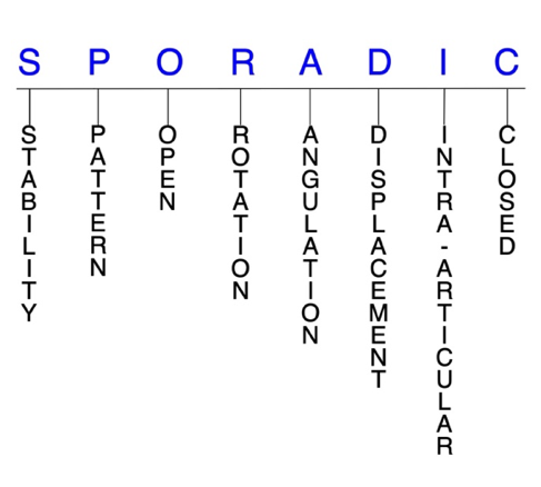
Hand Surgery Resource’s Diagnostic Guides describe fractures by the anatomical name of the fractured bone and then characterize the fracture by the Acronym:

In addition, anatomically named fractures are often also identified by specific eponyms or other special features. For the capitellar fracture, the historical and specifically named fractures include no common eponyms.
Fractures of the capitellum are coronal shear fractures of the distal humerus that rarely occur in isolation. The most common mechanisms of injury are falls on an outstretched hand (FOOSH) and falls that produce direct axial compression of the elbow while in a semi-flexed position. Associated bone and soft tissue injuries are extremely common and may include trochlear fractures, ligamentous injuries, ipsilateral fractures, and dislocations. Due to their low incidence, capitellar fractures are often overlooked during the initial evaluation, which can adversely affect long-term outcomes. Although conservative treatment with closed reduction and immobilization may be considered for minimally displaced capitellar fractures and displaced fractures without significant comminution, the general preference of most treating physicians is now surgical intervention.1-5
Definitions
- A capitellar fracture is a disruption of the mechanical integrity of the capitellum.
- A capitellar fracture produces a discontinuity in the capitellum contours that can be complete or incomplete.
- A capitellar fracture is caused by a direct force that exceeds the breaking point of the bone.
Hand Surgery Resource’s Fracture Description and Characterization Acronym
SPORADIC
S – Stability; P – Pattern; O – Open; R – Rotation; A – Angulation; D – Displacement; I – Intra-articular; C – Closed
S - Stability (stable or unstable)
- Universally accepted definitions of clinical fracture stability is not well defined in the literature.6-8
- Stable: fracture fragment pattern is generally nondisplaced or minimally displaced. It does not require reduction, and the fracture fragments’ alignment is maintained by with simple splinting or casting. However, most definitions define a stable fracture as one that will maintain anatomical alignment after a simple closed reduction and splinting. Some authors add that stable fractures remain aligned, even when adjacent joints are put to a partial range of motion (ROM). A stable capitellar fracture does not displace during active elbow motion.
- Unstable: will not remain anatomically or nearly anatomically aligned after a successful closed reduction and immobilization. Typically, unstable capitellar fractures have significant deformity with comminution, displacement, angulation, and/or shortening.
P - Pattern2
- Type 1: shear fracture involving most of the capitellum and little or none of the trochlea
- Type 2: variable amount of articular cartilage of the capitellum with minimal attached subchondral bone
- Type 3: comminuted or compression fracture of the capitellum
- Type 4: shear coronal fracture of the distal humerus involving the capitellum and most of the trochlea
O - Open
- Open: a wound connects the external environment to the fracture site. The wound provides a pathway for bacteria to reach and infect the fracture site. As a result, there is always a risk for chronic osteomyelitis. Therefore, open fractures of the capitellum require antibiotics with surgical irrigation and wound debridement.6,9,10
R - Rotation
- Capitellar fracture deformity can be caused by rotation of the proximal fracture fragment in relation to the distal fracture fragment.
- Degree of malrotation of the fracture fragments can be used to describe the fracture deformity.
- Fracture fragments in capitellar fractures are typically rotated internally.3
A - Angulation (fracture fragments in relationship to one another)
- Angulation is measured in degrees after identifying the direction of the apex of the angulation.
- Straight: no angulatory deformity
- Angulated: bent at the fracture site
D - Displacement (Contour)
- Displaced: disrupted cortical contours
- Nondisplaced: ≥1 fracture lines defining one or several fracture fragments; however, the external cortical contours are not significantly disrupted
- Fracture fragments in capitellar fractures are typically displaced proximally.3
I - Intra-articular involvement
- Intra-articular fractures are those that enter a joint with ≥1 of their fracture lines.
- All capitellar fractures are considered intra-articular fractures.3
- Isolated capitellar fractures can have fragment involvement with the radiocapitellar joint, while concomitant fractures with the trochlea can also involve the ulnohumeral joint.
- If a fracture line enters a joint but does not displace the articular surface of the joint, then it is unlikely that this fracture will predispose to post-traumatic osteoarthritis. If the articular surface is separated or there is a step-off in the articular surface, then the congruity of the joint will be compromised, and the risk of post-traumatic osteoarthritis increases significantly.
C - Closed
- Closed: no associated wounds; the external environment has no connection to the fracture site or any of the fracture fragments.4-6
Related Anatomy3,4,11,12
- The elbow is a complex hinge-type synovial joint comprised of the radius, ulna, and humerus, and formed by three articulations: the ulnohumeral joint, radiocapitellar joint, and proximal radioulnar joint.
- The ulnohumeral joint is a hinge joint in which the trochlear notch (or semilunar notch) of the ulna articulates with the trochlea of the humerus. This joint allows for elbow flexion and extension.
- The trochlea is the medial portion of the articular surface of the distal humerus, which is contained between the lateral and medial columns of the elbow and is primarily covered with articular cartilage. It has medial and lateral ridges with an intervening trochlear groove.
- The radiocapitellar joint is the articulation of the radial head with the capitellum of the humerus. It is essential to elbow longitudinal and valgus stability and has an integral relationship with the lateral collateral ligament (LCL).
- The capitellum is a smooth, round, hemispheric structure that represents a portion of a forward-and downward-projecting sphere and which forms the anterior and inferior articular surface of the distal humerus. It is covered with articular cartilage on its anterior and inferior sides, but not its posterior side.
- The elbow’s axis of flexion-extension and the axis of forearm rotation both pass through the capitellum, which enables effective reach by allowing the hand to function at different distances from the body.
- The key ligaments of the elbow include the lateral collateral ligament (LCL, which extends from the lateral epicondyle and blends with the annular ligament of the radius), the medial collateral ligament (MCL, which originates from the medial epicondyle and attaches to the coronoid process and olecranon of the ulna), and the annular ligament which encircles and stabilizes the radial head within the radial notch.
- The key tendons of the elbow include the tendons associated with the biceps, triceps and the extensor carpi radialis longus (ECRL) muscles as well as the common extensor tendon (the shared origin of the extensor carpi radialis brevis (ECRB), extensor digitorum communis (EDC), extensor digiti minimi (EDM) and extensor carpi ulnaris (ECU)), and the common flexor tendon (the shared origin of the pronator teres, flexor carpi radialis (FCR), palmaris longus, flexor digitorum superficialis (FDS), and flexor carpi ulnaris (FCU).
Incidence
- Capitellar fractures account for about 0.5–1% of all elbow fractures and about 6% of distal humerus fractures.3,12
- Capitellar fractures are more likely to occur in women than men, which may be related to cubitus valgus, cubitus recurvatum, or osteoporosis.12
- LCL injuries or radial head fractures have been documented in ~60% of patients following all coronal shear fractures of the distal humerus, which includes capitellar fractures.1
